Figures & data
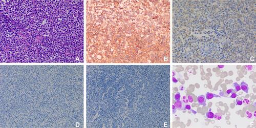
Figure 1 Ultrasound examination of the masses in the right breast and chest wall. (A) A heterogeneous hypoechoic mass with enhanced echo in the rear was found in the right breast. (B) A hypoechoic mass with striped hyperechoic feature was found in the chest wall. (C) Color Doppler Imaging of the mass in the chest wall displayed strip blood flow with high resistance index.
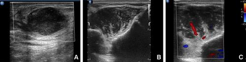
Figure 2 CT images of the masses in the right breast and chest wall. Before treatment (A–D): (A) There was a well-defined slightly high-density mass in the right breast (arrow). (B) An ill-defined iso-density mass ís shown in the chest wall (arrow). The masses in the breast (C) and the chest wall (D) show homogeneous and mild enhancement on enhanced CT images (arrow). After treatment (E and F): the masses of the breast (E) and chest wall (F) disappeared (arrow).
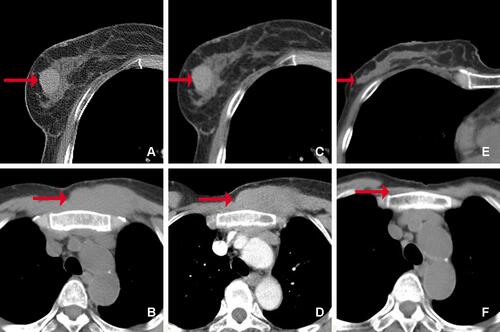
Figure 3 Histopathological examination of masses in the breast and chest wall. (A) HE staining shows lymphocyte proliferation (original magnification, ×200); immunohistochemical staining of the proliferated lymphocytes show positive for CD138 (B, original magnification, ×200) and IgD (C, original magnification, ×200), but negative for CD56 (D, original magnification, ×100) and CD117 (E, original magnification, ×100); (F) bone marrow smears (original magnification, ×1000) show proplasmocytes and immature plasma cells, which differ in size and shape. The cytoplasm was dark blue with a few granules. The nucleus is biased and binuclear with reticular chromatin and vacuoles in some nuclei and plasma.