Figures & data
Figure 1 Fundus photography. The swelling and blurry boundaries of both optic nerves were present (A and B). There was a splinter radial superficial retinal hemorrhage above the optic disc; no other obvious abnormality was observed (A and B). Three months after surgery, all these symptoms, including optic nerve swelling and retinal hemorrhage disappeared (C and D).
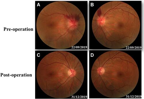
Figure 2 Brain MRI before surgery. Brain MRI showed a huge mass in the right temporal lobe, clear boundary, and multiple separations. The right temporal lobe and lateral ventricle were compressed, the midline structure shifted to the left (A and B). The lesion site showed low signal intensity on T1-weighted images (A) and high signal intensity on T2-weighted images (B). Contrast-enhanced T1-weighted images showed strong enhancement at the boundary (C and D).
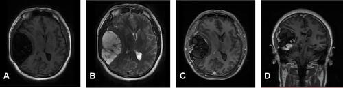
Figure 3 Surgical observations. The skull in the tumor area was partially protruding, the tumor invaded the skull, the adhesion of tumor tissue and skull was tight and the brain tissue of the right temporal lobe was compressed (A). The actual size of the tumor was 5.6 × 7.5 × 10.1 cm, and the texture was soft and gelatinous (B and C).
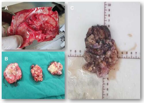
Figure 4 Hematoxylin and eosin staining. Foam cell accumulation in the mucous connective tissue of the right temporal lobe.
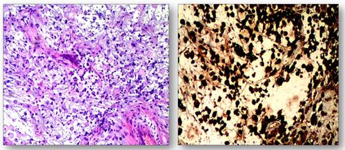
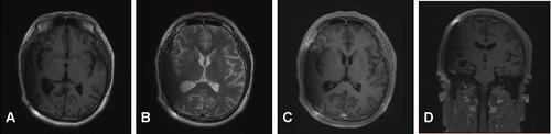
Figure 5 Three-month follow-up (MRI). T1-weighted and T2-weighted images show the midline structure returned to normal. A mild edema of temporoparietal was observed (A and B). Contrast-enhanced T1-weighted images (C and D) show no sign of recurrence.