
Figures & data
Figure 1 A 37-year-old female had severe sciatica for more than one year due to LDH and was treated by PELD using the transforaminal approach. (A–D) Preoperative X-rays and MR images showed protrusion at the L5-S1 segment. (E, F) Photograph during the operation showed the disc material removed and adequate decompression of the nerve root. (G–L) X-ray and MR images 7 years postoperation showed complete decompression and good preservation of the disc height.
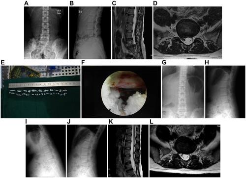
Table 1 Patient Demographic Data
Table 2 Clinical Outcomes Preoperative, 6 Months Postoperative and at Final Follow-Up