Figures & data
Figure 1 The result of CT-abdomen on March 11, 2019. (A) The invasion of SMA; (B) the metastasis of right liver; (C) the invasion of GDA and SMA; (D) the invasion of SMA; (E) the invasion of PV; (F) the invasion of SMV.
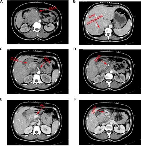
Figure 2 The histopathological result of percutaneous biopsy guided by CT. (A) 4×10 magnification; (B) 10×10 magnification; (C) 20×10 magnification.
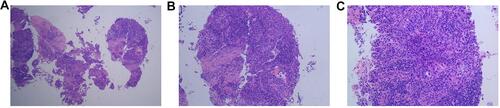
Figure 3 The X-ray image during TACE.
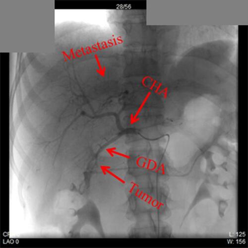
Figure 4 The result of CT-abdomen during the process of treatment. (A and D) The CT-abdomen result on April 15, 2019; (B and E) The CT-abdomen result on May 10, 2019; (C and F) The CT-abdomen result on July 26, 2019.
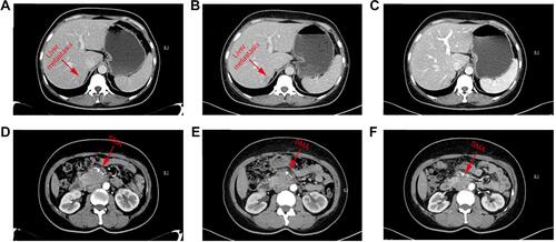
Figure 5 Changes of the tumor diameter and the main laboratory blood tests of this patient. (A) changes of the primary tumor; (B) changes of the liver metastasis; (C) changes of the CEA level; (D) changes of the CA125 level.
Abbreviation: PDCA, pancreatic ductal adenocarcinoma; CEA, carcinoembryonic antigen; CA125, carbohydrate antigen 125.
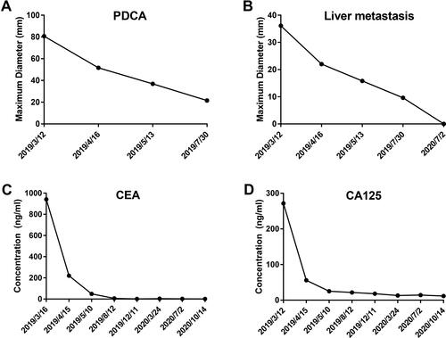
Figure 6 The postoperative specimen and histopathological result. (A) Postoperative specimen; (B) 4×10 magnification; (C) 10×10 magnification; (D) 20×10 magnification.
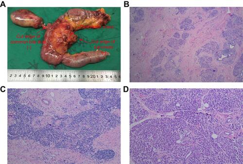
Table 1 Main Laboratory Test Results
Figure 7 The result of CT-abdomen after operation. There is no sign of recurrence of metastasis. (A and D) The CT-abdomen result on September 4, 2019; (B and E) the CT-abdomen result on March 23, 2020; (C and F) the CT-abdomen result on October 10, 2020.
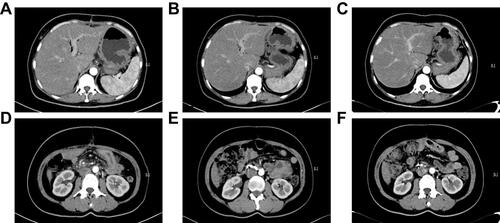
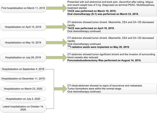
Figure 8 The flow chart of multidisciplinary treatment.