
Figures & data
Table 1 Baseline Characteristics of Subjects Classified According to Smoking Status
Figure 1 Image of irregular surface and calcification of carotid plaque for enrolled smokers and non-smokers, including CT scanning (left panel) and ultrasonography (right panel).
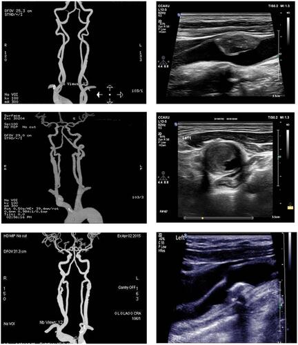
Table 2 Carotid Plaque Characteristics According to Smoking Status
Figure 2 Ridge trace analysis for different examination strategies. (A) Gold standard for plaque diagnosis. (B) Ultrasonography for determination of calcified plaque.
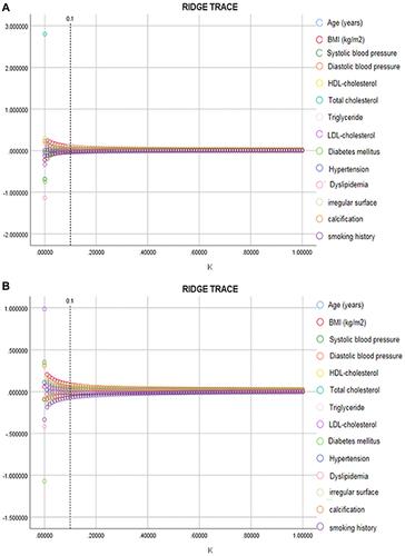
Figure 3 Analysis of plaque scores and age index, which were verified by ultrasonography index. X axis, age index; Y axis, scores for calcification and other plaque morphology. *P<0.05, **P<0.01.
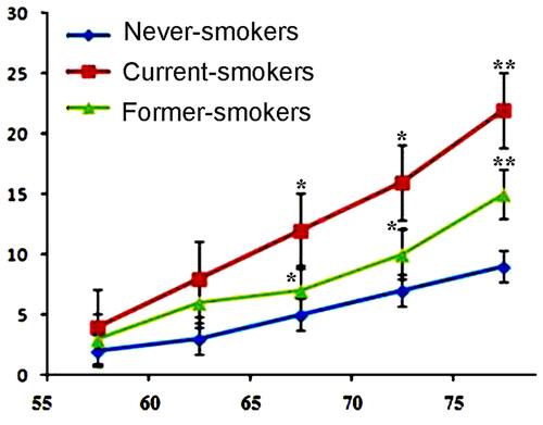
Table 3 Related Risk Factors Between Calcification and Plaque Surface Morphology