132
Views
1
CrossRef citations to date
0
Altmetric
Commentary

Identifying the Stroke Mimics

, ORCID Icon, & ORCID Icon
Pages 285-288 | Published online: 27 Jan 2021

Figures & data

Figure 1 Angioinvasive aspergillosis in a 20-year-old female with headache and acute onset aphasia. (A) T2W axial section shows inflammatory changes in the sphenoid sinus (white arrow) with altered signal intensity in the adjacent medial aspect of the left temporal lobe (black arrow). (B) Post contrast T1W axial section shows enhancing area in the region of left temporal lobe signal abnormality. (C) Diffusion study shows an acute infarct in the left temporal lobe related to infective vasculitis of left middle cerebral artery (white arrow). (D) Follow up T2W axial section after 15 months shows chronic infarcts in the left MCA territory.

Figure 1 Angioinvasive aspergillosis in a 20-year-old female with headache and acute onset aphasia. (A) T2W axial section shows inflammatory changes in the sphenoid sinus (white arrow) with altered signal intensity in the adjacent medial aspect of the left temporal lobe (black arrow). (B) Post contrast T1W axial section shows enhancing area in the region of left temporal lobe signal abnormality. (C) Diffusion study shows an acute infarct in the left temporal lobe related to infective vasculitis of left middle cerebral artery (white arrow). (D) Follow up T2W axial section after 15 months shows chronic infarcts in the left MCA territory.

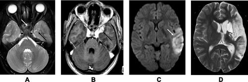
Table 1 Various Stroke Mimics Described in the Literature