Figures & data
Figure 1 The inferior retinal detachment with causative inferior breaks and multiple peripheral breaks that have been photocoagulated.
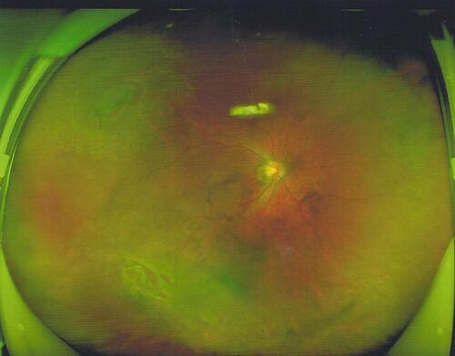
Figure 2 Retinal attachment on the 14th day postoperatively, with a small air bubble present in the vitreous cavity.
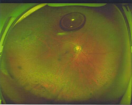
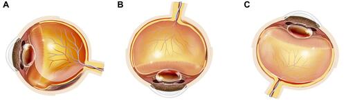
Figure 3 The schematic diagram showing the inferior breaks and the gas tamponade in different positions: (A) rhegmatogenous retinal detachment with causative inferior breaks. (B) Inferior retinal breaks not tamponaded in the prone position. (C) Inferior retinal breaks optimally tamponaded in the supine position.