Figures & data
Table 1 Demographic, Baseline and Clinical Characteristics of Patients Infected with COVID-19
Table 2 Laboratory Findings of Imported Patients Infected with COVID-19
Table 3 SARS-CoV-2 Nucleic Acid and Serum Anti-SARS-CoV-2 IgG and IgM Antibody Responses
Table 4 Chest CT Findings of Patients with COVID-19
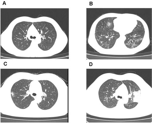
Figure 1 Chest CT images findings for four patients. (A) Chest computed tomography (CT) images of a 42-year-old male patient with COVID-19 taken on August 9, 2020, showing left lung pure GGO. (B) Chest CT images of a 54-year-old male patient with COVID-19 taken on August 9, 2020, showing GGO with reticular and interlobular septal thickening in both lungs. (C) Chest CT images of a 40-year-old female patient with COVID-19 taken on April 21, 2020, showing unilateral GGO with consolidation. (D) Chest CT images of a 33-year-old male patient with COVID-19 taken on April 27, 2020, showing left lung consolidation.