Figures & data
Table 1 Demographic Data and Clinical Risk Factors of Patients with Ruptured and Unruptured Basilar Tip Aneurysms
Table 2 BA Tip Aneurysm Characteristics Stratified by Rupture Status
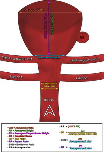
Figure 1 Stacked bar histograms illustrating the following: (A) patient demographics and aneurysmal hemodynamic status, and (B) morpho-radiological characteristics of the patient under investigation.
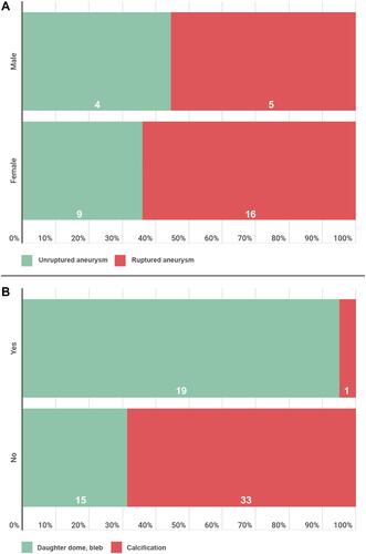
Figure 2 Ruptured basilar tip aneurysm. 3D CT angiography, (A) frontal and (B) lateral projections, shows a unilocular saccular aneurysm arising from the basilar tip (arrows) with a daughter sac at the left side of the aneurysm dome. The aneurysm is slightly inclined anteriorly. There is also an incidental finding of bilateral carotid cavernous fistulae.
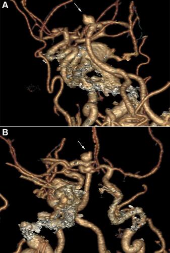
Figure 3 (A) Hemodynamically stable basilar tip aneurysm. The aneurysm is inclined posteriorly with smooth aneurysmal dome. (B) 3D lateral angiogram shows a unilocular saccular aneurysm arising from the basilar tip with posterior inclination (arrow).
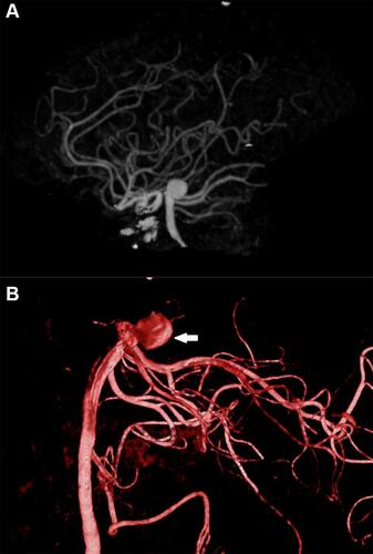
Figure 4 Schematic diagram illustrating calculation of BA aneurysm measurements.