
Figures & data
Figure 1 Endoscopic and endoscopic ultrasonography for gastrointestinal stromal tumor. (A) A submucosal tumor was detected in the gastric fundus by general gastroscopy. (B) Endoscopic ultrasonography showed that the lesion originated from the muscularis propria, was hypoechoic, and had a non-homogeneous echo and visible echo-free area; Doppler ultrasound did not identify blood flow; cross-section size: 2.0 cm * 2.5 cm.
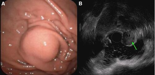
Table 1 Comparison of Five Surgical Methods for Gastric Stromal Tumors
Figure 2 Complete endoscopic full-thickness resection of tumor; clips combined with purse-string suture closure of wounds.
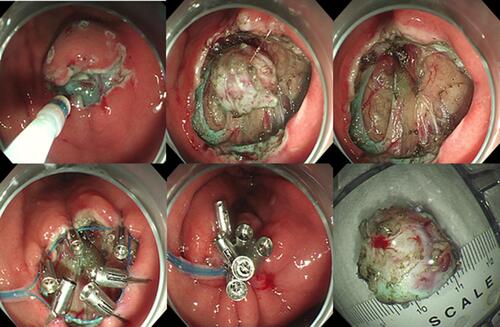
Table 2 Modified NIH Classification System Proposed by Joensuu
Figure 3 Electron microscope (× 40 [A], × 100 [B], × 200 [C]), hematoxylin and eosin staining of cross-sectioned gastrointestinal stromal tumor; spindle cells and mitotic figures can be seen.
![Figure 3 Electron microscope (× 40 [A], × 100 [B], × 200 [C]), hematoxylin and eosin staining of cross-sectioned gastrointestinal stromal tumor; spindle cells and mitotic figures can be seen.](/cms/asset/c6be6d4d-7586-4535-8108-f156e2fc283f/dijg_a_12168183_f0003_c.jpg)
Figure 4 Immunohistochemical staining of gastrointestinal stromal tumor with DOG1 (A), CD117 (B), CD34 (C).
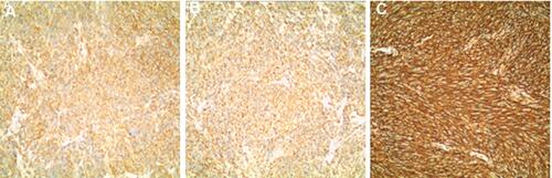
Table 3 Analysis of Ultrasound and General Endoscopic Features of Differentiated Gradient Stromal Tumors at Different Risks