Figures & data
Table 1 Demographic Features Between Two Groups Regarding Age, Weight, and Ejection Fraction
Table 2 NT-proBNP Levels and Oxidative Stress Markers in Asymptomatic and Symptomatic Patients
Table 3 Oxidative Stress Markers According to NT-proBNP Levels
Figure 1 Cardiac magnetic resonance and cardiac PET imaging (same patient). (A) In short axis view, no cardiac involvement on LGE images. (B) Increased 18F-FDG-PET uptake on lateral and inferolateral walls of left ventricle on bulls eye map and on slices (indicating jeopardized area).
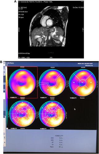
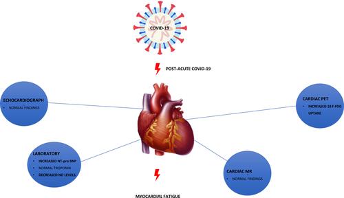
Figure 2 Effect of biochemical and imaging tests on possible cardiac involvement of post-acute COVID-19.