Figures & data
Figure 1 (A) Sonogram of nuchal cord clockwise coiling, the characteristic of blood flow: two red and one blue on the right half arc, and two blue and one red on the left half arc, it represents the umbilical cord coiling from right to left. (B) Sonogram of nuchal cord anticlockwise coiling, the characteristic of blood flow: two red and one blue on the left half arc, and two blue and one red on the right half arc, it represents the umbilical cord coiling from left to right.
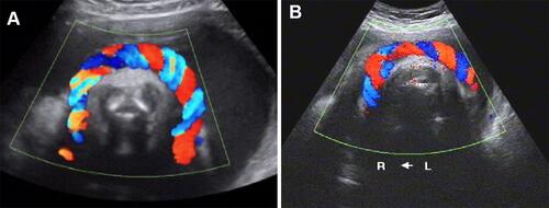
Figure 2 (A) α-shaped type: Left occipital transverse position, the spine is on the left side, the umbilical cord coils more than 1 cycle, 360° < angle < 540°; (B) O-shaped type: Anterior occipital position, the spine is in the front, the umbilical cord coils 1 cycle, the angle is 360°; (C) C-shaped type: Right occipital transverse position, the spine is on the right side, the umbilical cord coils less than 1 cycle, 180° < angle < 360°; (D) L-shaped type: Occipitoposterior position, the spine is at posterior site, the umbilical cord runs to the placenta through the anterior chest and one side of the neck, the coiling angle is ≤180°.
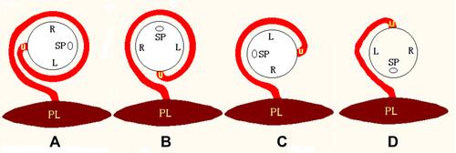
Figure 3 (A) α-shaped type: LOA position, right Figure shows the spine in the right front, the umbilical cord coils from left to right, left Figure shows the umbilical cord crossing the chest and entering the anterior wall of placenta, the coiling angle is >360°; (B) O-shaped type: The blood flow of the umbilical artery is in the direction of the arrow, the yellow arrow is the proximal end of the umbilical cord, and the green arrow is the distal end of the umbilical cord, the coiling angle is 360°; (C) C-shaped type: LOA position, the spine is on the left side, it can be seen that the umbilical cord coils from right to left, the distal end enters the posterior wall of placenta, the two ends of the umbilical cord are far away from each other and are hung in the neck like a scarf; (D) L-shaped type: LOA position, the spine is on the right side, the umbilical cord runs from the right anterior wall of the placenta through the left side of the fetal neck, the umbilical artery blood flow is blue, it represents the direction of the umbilical cord from the chest to the back.
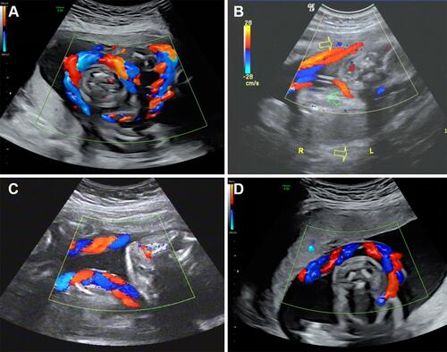
Figure 4 Type and direction of nuchal cord.
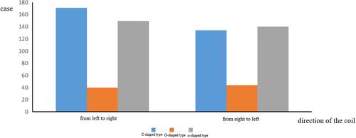
Figure 5 The relationship between the type of nuchal cord and placental position.
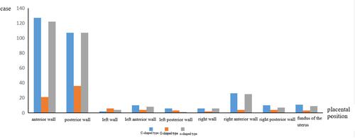
Figure 6 The relationship between the type of nuchal cord and spinal orientation.
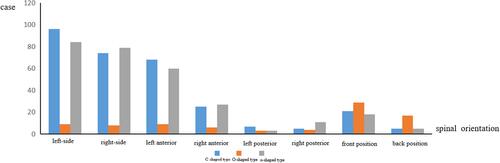
Figure 7 The relationship between coiling direction of nuchal cord and fetal position.
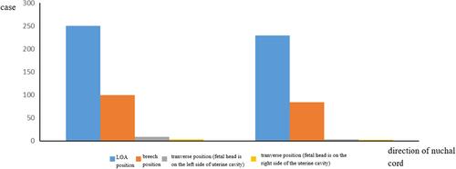
Figure 8 Model Figure of all types of nuchal cord.
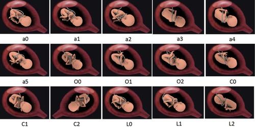
Figure 9 (A, B) If the direction of the blood flow is anti-clockwise, in case of LOA position, regardless of the spinal orientation, the umbilical cord coils from left to right; in case of breech presentation, regardless of the spinal orientation, the umbilical cord coils from right to left. (C, D) If the direction of the blood flow is clockwise, in case of LOA presentation, regardless of the spinal orientation, the umbilical cord coils from right to left; in case of in breech presentation, regardless of the spinal orientation, the umbilical cord coils from left to right.
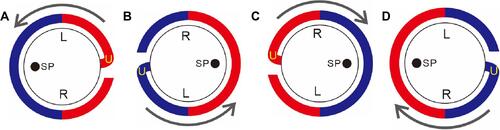
Figure 10 The relationship of the fetal position, placental location and the coiling direction of the umbilical cord with the coiling angle of the umbilical cord. When the coiling direction of the umbilical cord is the same (all from left to right or from right to left), but the spinal orientations are different, the coiling angles are different (A vs B and C vs D); when the coiling direction is different, and the spinal orientation is the same, the coiling angles are different (A vs C and B vs D); when the spinal orientations are different, and the coiling direction are different, the coiling angles are also the same (A vs D and B vs C); For population, even if the spinal orientations and the coiling directions are the same, but the placental locations are different, the coiling angles will also be different (E vs F). Therefore, the coiling angle of the umbilical cord is determined jointly by the placental location, the fetal position and the coiling direction of the umbilical cord.
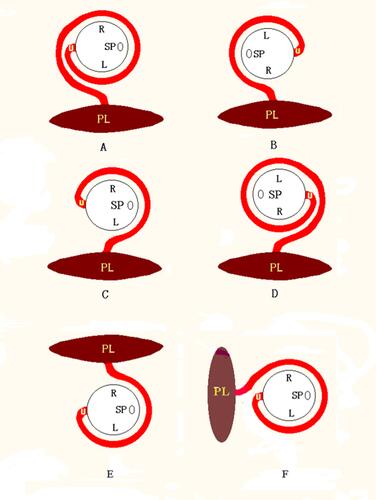
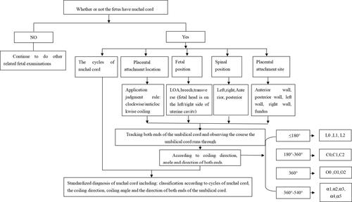
Figure 11 Standardized diagnosis process of nuchal cord.