
Figures & data
Table 1 The General Characteristics and Follow-Up Data in 15 Cases with Cervicothoracic Tuberculosis and Kyphosis
Figure 1 A female patient of 7 years old with C7-T9 vertebral tuberculosis and kyphosis. One-stage posterior approach internal fixation orthopedics was conducted followed by two-stage anterior approach lesion removal with additional bone grafting. (A and B) The preoperative whole spine frontal and lateral x-ray showed cervicothoracic segmental kyphosis, and obvious C7-T9 localized lateral kyphosis. (C) The preoperative MRI indicated the destruction of the C7~T9 vertebral body and intervertebral disc, a small amount of abscess formation in the paravertebral area, and a change in the intervertebral disc signal. (D and E) The preoperative CT and sagittal 3D reconstruction suggested multiple bone destruction at C7-T9, with some of the bone edges becoming hyperplastic and sharp. (F and G) The whole spine frontal and lateral x-ray after the staged posterior-anterior combined surgery showed a good position of the internal fixation and a good sequence of cervicothoracic vertebrae. (H and I) The whole spine frontal and lateral x-ray one year after surgery showed good internal fixation and complete osseous fusion.
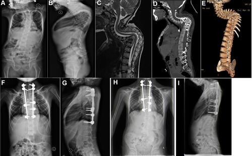
Figure 2 A female patient of 3 years old with C4–T4 vertebral tuberculosis and kyphosis. One-stage posterior approach internal fixation orthopedics was conducted followed by two-stage anterior approach lesion removal with additional bone grafting. (A and B) The preoperative whole spine frontal and lateral x-ray showed obvious C4–T4 kyphosis. (C) The preoperative MRI showed destruction of C4~T4 vertebral body and intervertebral disc, with paravertebral abscess formation and change in the intervertebral disc signal. (D and E) The preoperative CT and sagittal 3D reco nstruction suggested C4-T4 multiple arch bone destruction with C6 vertebral body collapse. (F and G) The whole spine frontal and lateral x-ray after the staged posterior-anterior combined surgery showed a good position of the internal fixation. (H and I) The whole spine frontal and lateral x-ray six year after surgery showed good internal fixation and complete osseous fusion.
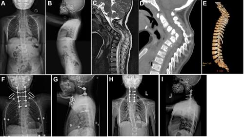
Table 2 Results of Imaging Measurements Before and After Surgery and at the Last Follow-Up in 15 Pediatric Patients
Table 3 The Results of JOA Score, VAS, and NDI Before and After Surgery and at the Last Follow-Up in 15 Pediatric Patients