Figures & data
Table 1 Location of Giant Cell Tumors (GCTBs) in 57 Patients 18 Years of Age and Younger
Figure 1 The distribution of age in 57 patients with giant cell tumors 18 years of age and younger.
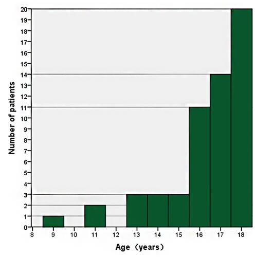
Figure 2 A skeletally mature 16-year-old girl with GCTB in the distal femur. Plain radiographs (A and B) of the knee demonstrate an eccentric expansile osteolytic lesion involving the distal femoral epiphysis and metaphysis. The lesion is well defined with a narrow zone of transition (arrow). CT (C and D) showed focal loss of bone cortex and adjacent soft tissue extension (arrow).
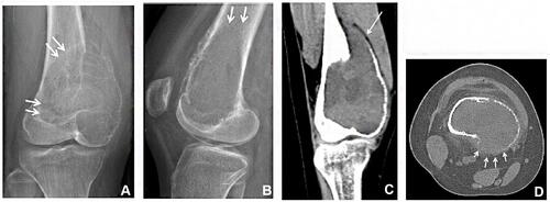
Table 2 Patient Characteristics and Imaging Features of Pediatric (18 Years of Age and Younger) and Adult (Over 18 Years) Patients with GCTB Occurring in Long Tubular Bones
Table 3 Evaluation of Skeletal Maturity of GCTB Patients 18 Years of Age and Younger
Table 4 GCTBs in Patients 18 Years of Age and Younger: Extent of Involvement of the Lesions in Long and Short Tubular Bones
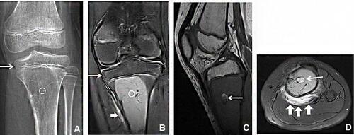
Figure 3 A skeletally immature 9-year-old girl with GCTB in the proximal tibia. Plain radiograph (A) demonstrates an osteolytic lesion (white circle) centered in the metadiaphysis, sparing the epiphysis (arrow in a). Coronal fat-suppressed T2 (B), sagittal T1 (C), and axial fat-suppressed T2 (D) show a solid lesion (white circle) in the proximal tibial metadiaphysis with sparing of the epiphysis (arrow in b). The lesions were mainly solid, with a few cystic components (arrows in c and d). Adjacent soft tissue edema were shown on coronal and axial T2WI (thick arrows in b and d).