
Figures & data
Figure 1 Representative SWE images of the hardest color. The hardest color, which corresponds to the maximum hardness inside a nodule, includes blue (A), green (B), Orange (C), and red (D).
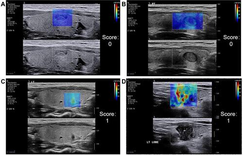
Figure 2 Representative SWE images of the main color. The main color, which fills the entire nodule or is predominant in the nodule, includes blue (A), green (B), Orange (C) and red (D).
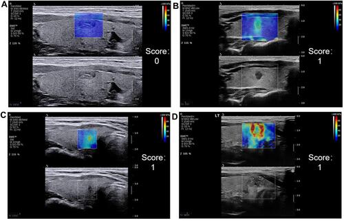
Figure 3 Representative SWE images of the stiff rim. The stiff rim is a high-hardness area with an annular or semi-cyclic distribution at the edge of a nodule. Representative SWE images with stiff rim (A) and without stiff rim (B).
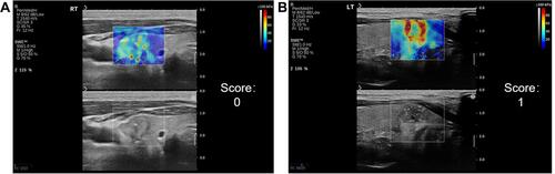
Figure 4 Representative SWE images of internal color homogeneity. Homogeneity refers to the appearance of only one color in a nodule (A), while inhomogeneity refers to the appearance of two or more colors in a nodule (B).
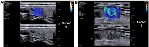
Figure 5 Representative SWE images of color homogeneity with surrounding glands. Homogeneity refers to the consistency of the color of the nodule with its surrounding glands (A), while inhomogeneity refers to the inconsistency of the color of the nodule with its surrounding glands, showing a clear edge to separate the nodule from its surrounding glands (B).
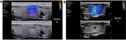
Table 1 Baseline Information of Patients with Malignant and Benign Thyroid Nodules
Table 2 SWE Color Characteristics for Benign and Malignant Nodules
Table 3 Malignancy Rates and SWE Color Scores
Figure 6 SWE images and HE staining of benign thyroid nodule. Imaging with SWE (A) shows blue as the hardest color, blue as the main color, no stiff rim, homogeneity of the internal color, and the color between the nodules and its surrounding. The SWE score was 0. HE histopathological staining (magnification, 40x). Nodular goiter: Follicles vary in size and are nodular. Imaging with SWE (B) shows green as the hardest color, blue as the main color, no stiff rim, inhomogeneity of the internal color, and the color between the nodules and its surrounding. The SWE score was 1. HE histopathological staining (magnification, 100x). Adenomatous goiter: a single nodule with a capsule visible on the surface. The follicles in the capsule are relatively uniform in size, showing adenoma-like hyperplasia.
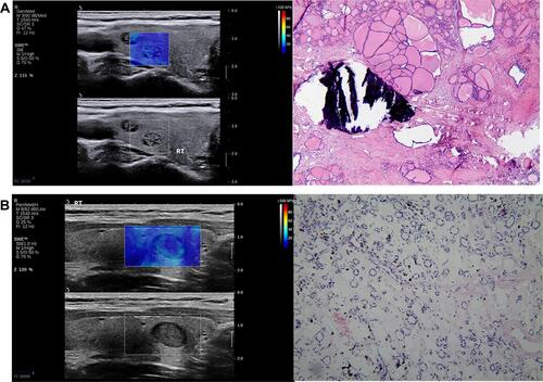
Figure 7 SWE images and HE staining of malignant thyroid nodule. Imaging with SWE (A) shows red as the hardest color, green as the main color, no stiff rim, inhomogeneity of the internal color, and the color between the nodules and its surrounding. The SWE score was 4. HE histopathological staining (magnification, 40x). Papillary microcarcinoma: The tumor grows liked a papillary, with enlarged nuclei, crowded, ground glass-like, with nuclear grooves; interstitial fibrous tissue hyperplasia and hyaline degeneration. Imaging with SWE (B) shows red as the hardest color, green as the main color, showing stiff rim, inhomogeneity of the internal color, and the color between the nodules and its surrounding. The SWE score was 5. HE histopathological staining (magnification, 100x). Papillary carcinoma: The tumor was papillary and follicular growth, the nucleus was enlarged, deeply stained, crowded, ground glass-like, and nuclear grooves could be seen; interstitial fibrous tissue hyperplasia, hyaline degeneration.
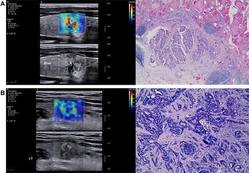
Table 4 Diagnostic Performance of Different Parameters Analyzed by Receiver Operating Characteristic (ROC) Curves Analysis
Table 5 Reliability Between Clinicians