
Figures & data
Table 1 Age, Gender and Lesion Side Distribution of Included Patients
Figure 1 (A) Posterior auricular skin flap preparation; (B) a transfer of the posterior auricular skin flap to the front of the ear; (C) repair of the preauricular defect with the posterior auricular skin flap; (D) surgical site appearance after the flap suture.
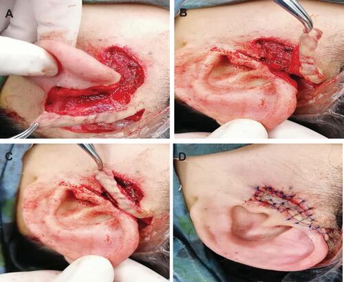
Figure 2 (A) Pre-operation label of the surgical incision; (B) resection of the preauricular lesion; (C) preparation of the Burow flap; (D) surgical site appearance after the preauricular incision is sutured.
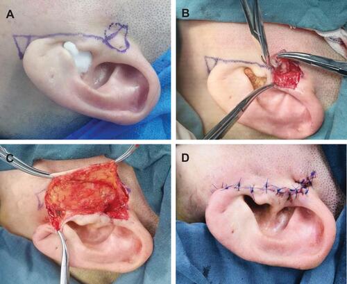
Table 2 Treatment Duration and Total Cost
Table 3 Analysis of Outpatient Medicine/Treatment Cost and Inpatient Medicine/Treatment Cost in Conventional and Local Flap Repairing Treatment Groups
Table 4 Post-Operation Complications