
Figures & data
Figure 1 Optical coherence tomography of vitreomacular traction. That is by definition a structural abnormality, which causes focal, tractional, distortion of the macula.
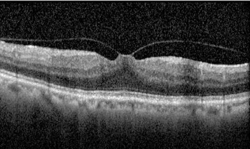
Table 1 Factors Associated with Vitreous Macular Traction in the Beijing Eye Study (Univariate Analysis)
Figure 2 Diagram showing the distribution of prevalence of vitreous macular traction stratified by age groups in Beijing Eye Study 2011.
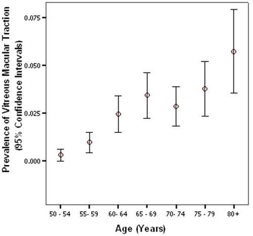
Table 2 Factors Associated with Vitreous Macular Traction Using Multivariate Logistic Regression Models in the Beijing Eye Study