Figures & data
Figure 1 ECG of a 46-year-old man with chest pain for 1 hour at the time of admission. Leads II, III, and AVF show a slur-type ischaemic J wave (indicated by red arrows). CAG showed the culprit artery to be the LAD, which was consistent with the leads showing an ischaemic J wave. Twenty-seven minutes later, an ischaemic J wave in leads II and III and AVF disappeared, indicating that the J wave occurred in the very early phase and changed with the development of ischaemia.
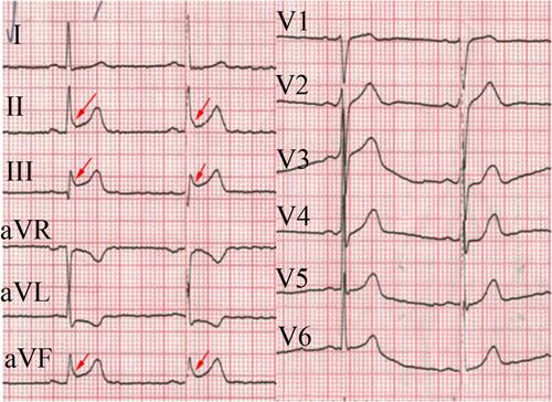
Figure 2 The ECG of a 55-year-old man presenting with chest pain for approximately 1 h. A notch J wave (indicated by red arrows) is present in leads II, III, AVF, and V6 with ST segment elevation consistent with the scope of blood supply to the RCA.
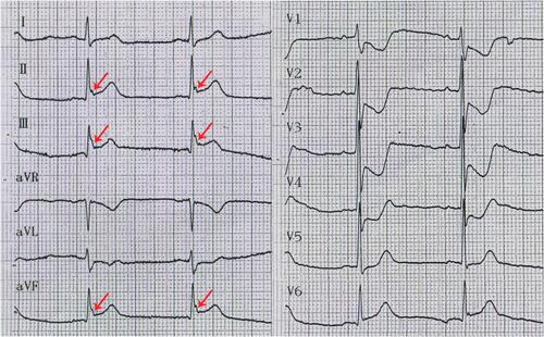
Figure 3 A BrP in leads V1-V4 and sudden cardiac death occurred in this patient. A BrP with ischaemic J wave is indicated by the red arrows.
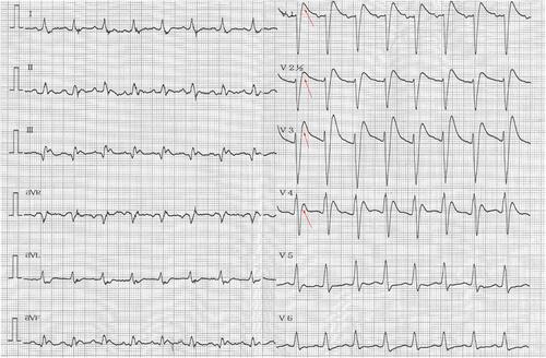
Table 1 Characteristics of STEMI Patients
Table 2 Univariate and Multivariate Logistic Regression Analyses of Ventricular Fibrillation Occurrence
Table 3 Univariate and Multivariate Logistic Regression Analyses of Ventricular Fibrillation
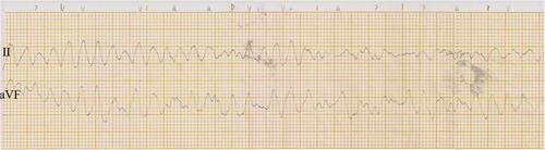
Figure 4 The records for the patient are shown in . When he returned to the CCU after the procedure, ventricular arrhythmia was noted, and he was shocked immediately. He declined an ICD implant and requested discharge 7 days later.