Figures & data
Table 1 Frequency of Different Tumors Stained with TLE1 Antibody (n = 177)
Table 2 Comparison of Frequency of Overall TLE1 IHC Expression in Synovial Sarcoma and Non-Synovial Sarcoma Cases (n = 177)
Table 3 Comparison of Frequency of Intensity of TLE1 IHC Expression in Synovial Sarcoma and Non-Synovial Sarcoma Cases (n = 177)
Figure 1 TLE1 IHC staining in synovial sarcoma and Ewing sarcoma. (A) Poorly differentiated synovial sarcoma, H&E, 20x. (B) Strong positive nuclear TLE1 IHC expression in tumor cells, 20x. (C) Ewing sarcoma, H&E, 20x. (D) Negative TLE1 IHC expression in cells of Ewing sarcoma tumor cells, 20x.
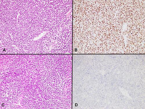
Table 4 Summary of Intensity-Wise TLE1 Expression in Non-Synovial Sarcoma Entities Showing Positive TLE1 Expression
Figure 2 TLE1 IHC staining in cellular schwannoma and leiomyosarcoma. (A) Cellular schwannoma, H&E, 20x. (B) Strong nuclear TLE1 expression in tumor cells of cellular schwannoma, 20x. (C) Leiomyosarcoma, H&E, 20x. (D) Moderate nuclear TLE1 expression in tumor cells of leiomyosarcoma, 20x.
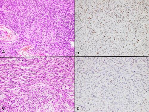
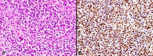
Figure 3 TLE1 staining in malignant melanoma. (A) Malignant melanoma, H&E, 20x. (B) Strong nuclear expression of TLE1 IHC stain in melanoma cells, 20x.