Figures & data
Table 1 Clinical Characteristics of Post-COVID-19 Patients
Table 2 Ocular Findings on Eye Examination of Patients Post-COVID-19
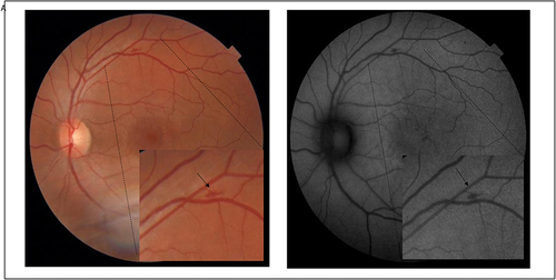
Figure 1 Main retinal findings and OCT follow-up patient 1 (A–C). (A) Colour fundus photography and red-free 30-days after symptoms imaging of the right eye of a female patient in her 60s revealing well-delimited sectorial pallor at the lower temporal arcade (black arrow). Vasoactive pharmacological support was not necessary during hospitalisation. (B) Colour fundus photography and red-free 70-days with disappearance of the lesion. (C) OCT-A 30-day follow-up shows a triangular area of low blood flow (white arrowhead), corresponding to the region of retinal pallor. (D) OCT B-scan in the 70-day follow-up of the retinal pallor. Note the local retinal thinning, with loss of differentiation of the inner layers of the retina (white arrowhead).
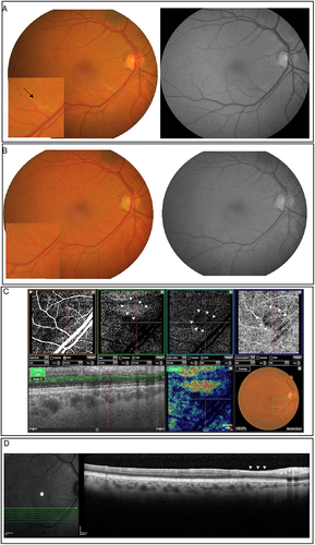
Figure 2 Main retinal findings and OCT follow-up patient 2 (A–C). (A) Colour fundus photography and red-free imaging of the left eye of a male patient revealing a peripapillary and macular flame-shaped haemorrhage (black arrow). Vasoactive pharmacological support was not necessary during hospitalisation. (B) Intraretinal cysts in the outer nuclear layer with 41-day after symptoms. (C) OCT 68-days after symptoms without intraretinal cysts.
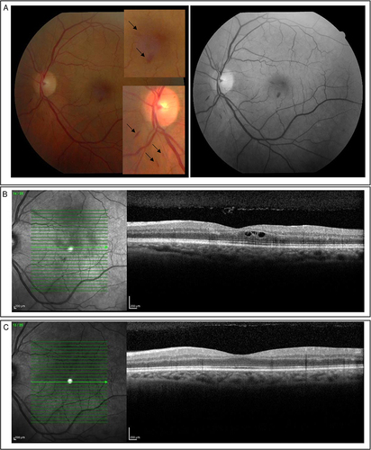
Figure 3 Main retinal findings patient 3 (A) Colour fundus photography and red-free imaging of the left eye of a male patient, without comorbidities and revealing a flame-shaped haemorrhage (black arrow). He had a mild condition, without hospitalisation.