Figures & data
Figure 1 Ultrasound of a 25-year-old woman with a ruptured epidermal inclusion cyst in the left leg shows a well-circumscribed subcutaneous cystic mass with evidence of wall rupture and leakage of turbid content to the surrounding tissue.
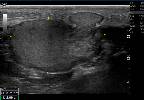
Figure 2 Ultrasound of a 30-year-old woman with cervical nodal enlargement shows an enlarged lymph node with hilar hypervascularity. Lymph node biopsy revealed Kikuchi histiocytic necrotizing lymphadenitis.
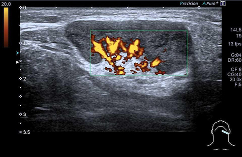
Figure 3 Ultrasound of a 35-year-old man with a wooden foreign body and abscess formation in the right leg shows an elongated hyperechoic structure representing the wooden foreign body within the thick-walled fluid collection in the subcutaneous layer of the right leg.
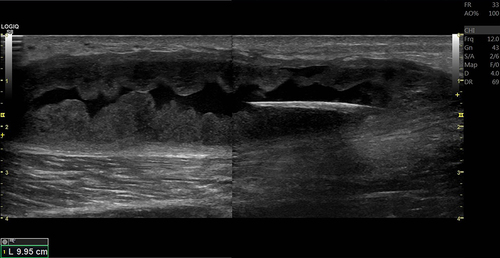
Figure 4 Ultrasound of a 41-year-old man with a lump on his chin shows a subcutaneous cyst with a scolex at the anterior wall. An inflammatory reaction is seen around the cyst. The pathology confirmed the diagnosis of cysticercosis.
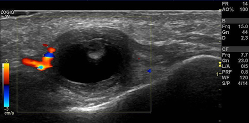
Figure 5 Doppler ultrasound of a 1-day-old newborn with a bluish subcutaneous nodule in the anterior abdominal wall shows a well-circumscribed hypoechoic lesion in the subcutaneous plane with internal hypervascularity, compatible with soft tissue hemangioma.
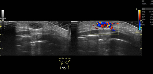
Figure 6 Doppler ultrasound of a 55-year-old man with superficial thrombophlebitis caused by a spider bite shows an intraluminal thrombus in the dilated lumen and thickened wall of the great saphenous vein of the left leg.
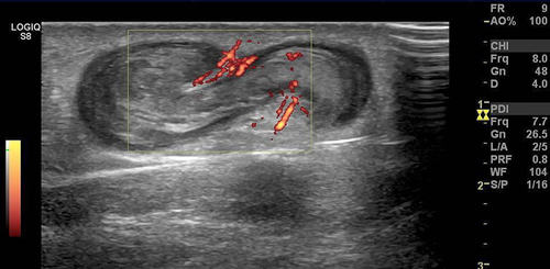
Figure 7 High-frame rate vector flow imaging of the knee was performed to exclude deep vein thrombosis. The image shows normal patency of the popliteal vein with green vector arrows. No thrombosis is found.
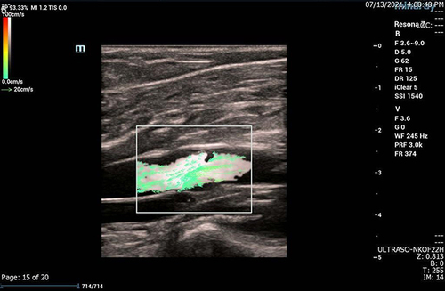
Figure 8 Ultrasound elastography of a 50-year-old woman with invasive ductal carcinoma of the right breast shows that the cancer mass has lower elasticity (red) than the surrounding normal breast tissue (yellow, green, and blue).
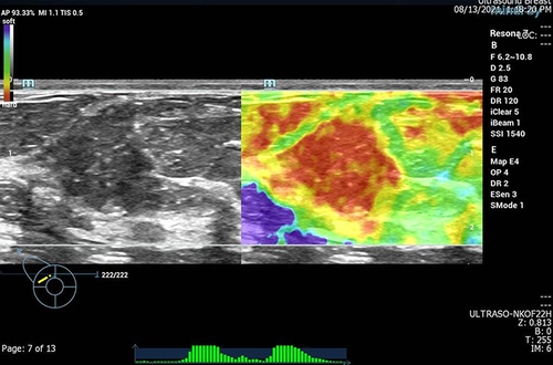
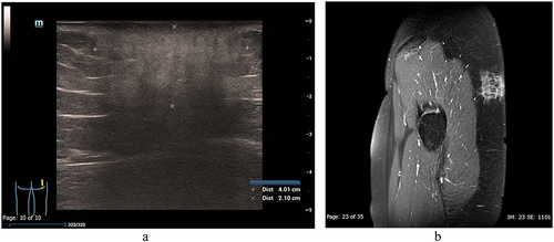
Figure 9 (a and b) A 21-year-old woman with pathologically proven lupus panniculitis on her right buttock. (a) Ultrasound shows the hyperechoic lesion with vague and ill-defined borders involving the subcutaneous fat, representing the inflammation of the fat. (b) T1-weighted fat-saturated gadolinium-enhanced MRI, sagittal view, shows the enhanced infiltrating lesion involving the skin and subcutaneous fat. The underlying muscle is preserved. MRI can delineate the deep margins of the lesion.