Figures & data
Figure 1 Fully automated hybrid model for HE prediction. In Model 2, we use feature-level fusion approaches for fusion of features with the AIM of collecting complementary information from radiomics, clinical data.
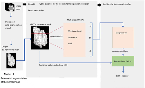
Table 1 The Characteristics of All Inclusion Participants
Figure 2 The patient enrollment process.
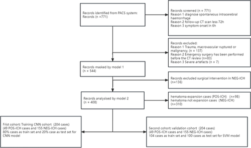
Figure 3 Representative image of hematoma by automatically labeled. The effect of the automatic hematoma labeling tool on a certain patient. The red color in the above picture shows the range of the labeled hematoma, and the picture below shows the original axial image of the brain.
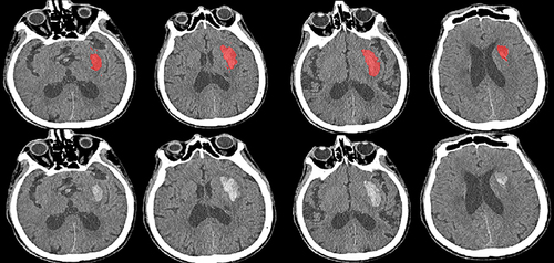
Table 2 Results of Hybrid Module Receiver Operating Characteristic, Precision-Recall
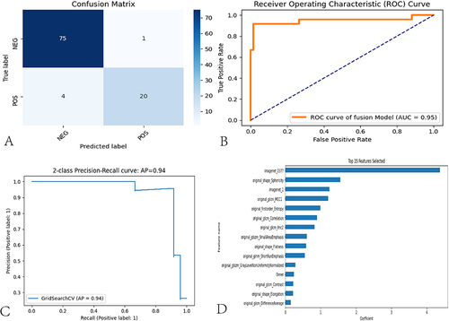
Figure 4 Performance of the hybrid model in the prediction of HE status. (A) The confusion matrix shows how well the model predicts the test set. (B) ROC curve of the model. (C) The precision-recall curve of the model. (D) Top 15 features with high importance in the SVM based classifier.