Figures & data
Table 1 Baseline Clinical Characteristics of 33 Patients
Table 2 Radiologic, and Histopathologic Tests
Figure 1 Chest CT results of TO demonstrated calcified nodules in the anterolateral wall protruding into the tracheal lumen. (A) Calcified nodules in the anterior wall (arrow). (B) Calcified nodules in the lateral wall (arrow).
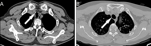
Figure 2 Bronchoscopic results of TO in different stages. (A) Stage I: Scattered nodular protrusions in the lower part of the trachea near the bulge; (B) Stage II: Diffuse distribution of nodular protrusions on the anterior and lateral walls of the trachea; (C and D) Stage III: Deformation and narrowing of the lumen.
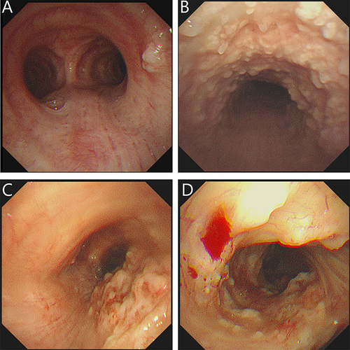
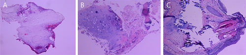
Figure 3 Histopathological findings of TO illustrateTracheal mucosa intact. (A) Nodular cartilaginous tissue with calcification; (B) Nodular cartilaginous tissue and mature bone tissue with calcification; (C) Squamous epithelial metaplasia of the tracheal mucosa and nodular mature cartilage and bone tissue were seen in the interstitium.