Figures & data
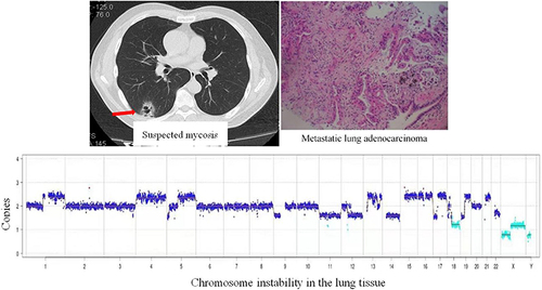
Table 1 Comparison of General Data Between LRTIs and Non-LRTIs
Figure 1 Flow diagram showing patient enrollment. aUncertain pathogen, it is unclear whether the detected pathogen is pathogenic or colonizing bacteria; bRespiratory tract specimens, including sputum, alveolar lavage fluid, and lung biopsy tissue.
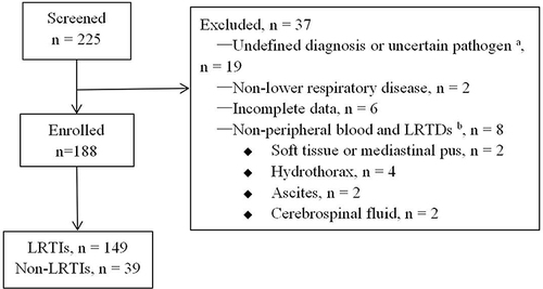
Table 2 Comparison of the Pathogen Detection Techniques
Table 3 Comparison of Detection Rates of Different Pathogen Types Between mNGS and CMT
Table 4 Clinical Data of 15 Cases of Malignant Tumor with CNVs and Pathogens Detected by mNGS
Figure 2 (A) Chest computed tomography scan showing multiple pulmonary bullae, multiple patchy and large flake consolidation in both lungs, with predominance in the lower lobes. (B) Electronic gastroscopy revealing an irregular mass in the fundus of the stomach involving the upper body of the stomach. (C) Stomach biopsy revealing poorly differentiated adenocarcinoma. (D) Biopsy of the left lower lobe of the lung revealing invasive adenocarcinoma, originating from the stomach.
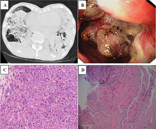
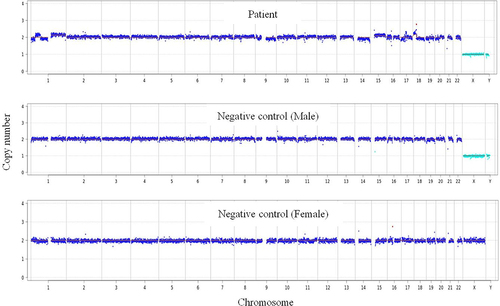
Figure 3 Chromosome detection revealing genome instability due to CNV, indicating a highly suspected tumor.