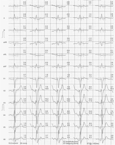
Figures & data
Figure 1 ECG at rest from brother A.
Abbreviations: aVF, augmented unipolar limb lead in which the positive electrode is on the left leg; aVL, augmented unipolar limb lead in which the positive electrode is on the left arm; aVR, augmented unipolar limb lead in which the positive electrode is on the right arm; ECG, electrocardiogram; SBS, SCHILLER Baseline-Stabilizer.
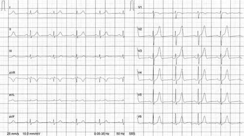
Figure 2 Exercise ECG from brother A.
Notes: First row = at rest; second row = last step before maximum exertion; third row = maximum exertion; fourth row = after 2 minutes of recovery; fifth row = after 4 minutes of recovery.
Abbreviations: aVF, augmented unipolar limb lead in which the positive electrode is on the left leg; aVL, augmented unipolar limb lead in which the positive electrode is on the left arm; aVR, augmented unipolar limb lead in which the positive electrode is on the right arm; ECG, electrocardiogram.
Abbreviations: aVF, augmented unipolar limb lead in which the positive electrode is on the left leg; aVL, augmented unipolar limb lead in which the positive electrode is on the left arm; aVR, augmented unipolar limb lead in which the positive electrode is on the right arm; ECG, electrocardiogram.
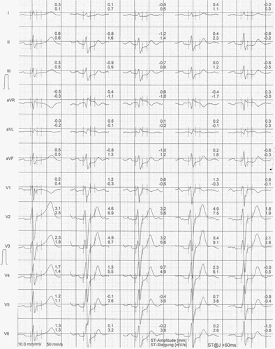
Table 1 Laboratory results of the two brothers at the time of the checkup
Figure 3 ECG at rest from brother B.
Abbreviations: aVF, augmented unipolar limb lead in which the positive electrode is on the left leg; aVL, augmented unipolar limb lead in which the positive electrode is on the left arm; aVR, augmented unipolar limb lead in which the positive electrode is on the right arm; ECG, electrocardiogram; SBS, SCHILLER Baseline-Stabilizer.
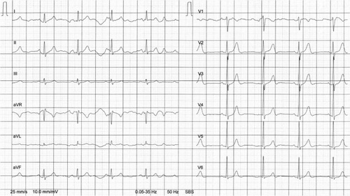
Figure 4 Exercise ECG from brother B.
Notes: First row = at rest; second row = last step before maximum exertion; third row = maximum exertion; fourth row = after 2 minutes of recovery; fifth row = after 4 minutes of recovery.
Abbreviations: aVF, augmented unipolar limb lead in which the positive electrode is on the left leg; aVL, augmented unipolar limb lead in which the positive electrode is on the left arm; aVR, augmented unipolar limb lead in which the positive electrode is on the right arm; ECG, electrocardiogram.
Abbreviations: aVF, augmented unipolar limb lead in which the positive electrode is on the left leg; aVL, augmented unipolar limb lead in which the positive electrode is on the left arm; aVR, augmented unipolar limb lead in which the positive electrode is on the right arm; ECG, electrocardiogram.