Figures & data
Figure 1 TNF-α-initiated signal transduction. TNF-α (soluble or membrane-bound) activates two different TNF-receptors, ie, TNFR1 and TNFR2. After binding of TNF-α, TNFR1 recruits distinct adaptor molecules (TRAF2, TRADD) at the intracellular death domain, thereby activating three major signaling pathways: NF-κB-signaling, MAPK/C-Jun-signaling, and caspase/apoptotic signaling. TNFR2 activation by membrane-bound TNF-α leads to activation of PI3K/Akt and proangiogenic pathways (eg, VEGF/VEGFR2), and is furthermore able to interfere with NF-κB signaling.
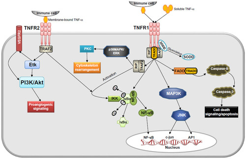
Figure 2 Shear stress dependency of TNF-α signaling in endothelial cells. (A) Laminar shear stress prevents proinflammatory activation of endothelial cells by TNF-α by inhibiting NF-κB signaling (eg, preventing expression of adhesion molecules and inducing production of NO) and therefore results in a quiescent endothelial phenotype. (B) Proatherogenic non-uniform shear stress synergistically enhances proinflammatory TNF-α signaling by activating NF-κB, AP-1, or c-Jun, leading to endothelial dysfunction characterized by increased permeability, enhanced oxidative stress, lack of cell alignment with flow and adhesion molecule expression, followed by adhesion of leukocytes to the activated endothelium.
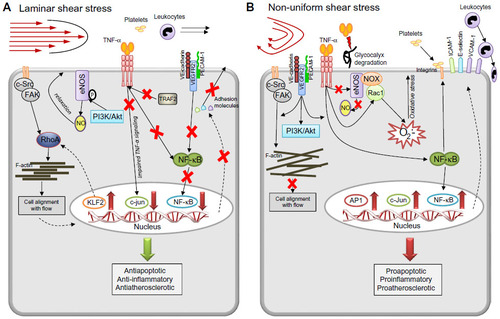
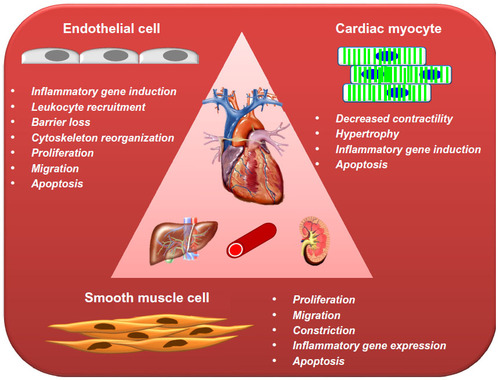
Figure 3 Schematic presentation of the most important effects elicited by TNF-α in the cells of cardiovascular system.