Figures & data
Table 1 Hydrodynamic radii (dH), ζ-potential, and polydispersity index (PDI) of different superparamagnetic iron oxide nanoparticles (SPIONs): uncoated (Un), coated with D-mannose (Man), or poly-L-lysine (PLL) in ultrapure water (UPW) and cell-free culture medium (DMEM) after 1 hour
Table 2 Evaluation of primary DNA damage in neural stem cells following 24-hour exposure to uncoated superparamagnetic iron oxide nanoparticles (UnSPIONs), and coated with d-mannose (ManSPIONs) or poly-L-lysine (PLLSPIONs)Table Footnotea
Figure 1 Transmission electron microscopy of differently coated superparamagnetic iron oxide nanoparticles.
Notes: (A) Uncoated; (B) coated with D-mannose; (C) coated with poly-L-lysine. Images were recorded at ×85,000 magnification.
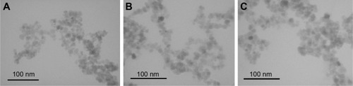
Figure 2 Effect of superparamagnetic iron oxide nanoparticles (SPIONs) with different surface coating on cell viability measured by the Cell Counting Kit 8 assay.
Notes: Neural stem cells were exposed to different concentrations, given in mg/L, of SPIONs for 24 hours. Control cells were cultivated in nanoparticle-free exposure media (negative controls) or treated with dimethyl sulfoxide (positive controls). The data for cell viability, expressed as the mean of three independent experiments conducted in five replicates, were calculated as percentages of the values measured in control cells. Error bars represent standard deviation. *P<0.05 compared with negative control.
Abbreviations: UnSPIONs, uncoated SPIONs; ManSPIONs, D-mannose-coated SPIONs; PLLSPIONs, poly-L-lysine-coated SPIONs.
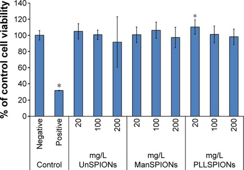
Figure 3 Effect of SPIONs with different surface coating on ROS levels measured by the DCFH-DA and DHE staining methods.
Notes: Neural stem cells were exposed to different concentrations of SPIONs, given in mg/L, for 4 hours (solid filled columns). To evaluate possible neural stem cell recovery from acute oxidative stress, cells were placed in fresh nanoparticle-free Dulbecco’s Modified Eagle’s Medium for an additional 4 hours after exposure to SPIONs (dotted columns). Control cells were cultivated in nanoparticle-free exposure media (negative controls) or treated with 100 μM of hydrogen peroxide (positive controls). The data for cell viability, expressed as the mean of three independent experiments conducted in five replicates, were calculated as percentages of the values measured in control cells. Error bars represent standard deviation. *P<0.05 compared with negative control.
Abbreviations: SPIONs, superparamagnetic iron oxide nanoparticles; ROS, reactive oxygen species; DCFH-DA, dichlorodihydrofluorescein diacetate; DHE, dihydroethidium; UnSPIONs, uncoated SPIONs; ManSPIONs, D-mannose-coated SPIONs; PLLSPIONs, poly-L-lysine-coated SPIONs.
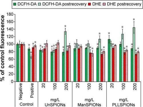
Figure 4 Effect of SPIONs with different surface coating on total GSH content measured by MBCl assay in neural stem cells.
Notes: After 4 hours of exposure (solid filled columns) and after 4 hours of recovery (dotted columns). The data, expressed as the mean of three independent experiments conducted in five replicates, were calculated as percentages of the values measured in negative controls (cells in nanoparticle-free exposure media). Positive controls represent cells treated with 100 μM of hydrogen peroxide. Error bars represent standard deviation. *P<0.05 compared with negative control.
Abbreviations: SPIONs, superparamagnetic iron oxide nanoparticles; GSH, glutathione; MBCl, monochlorobimane; UnSPIONs, uncoated SPIONs; ManSPIONs, D-mannose-coated SPIONs; PLLSPIONs, poly-L-lysine-coated SPIONs.
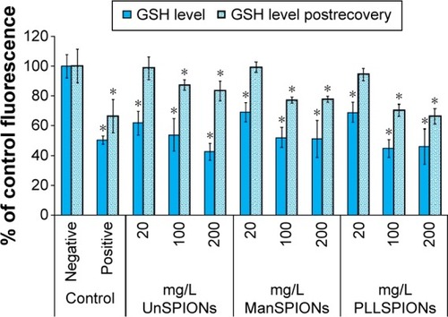
Figure 5 Effect of SPIONs with different surface coating on the activities of GPx and SOD in neural stem cells after 4 hours of exposure.
Notes: The data, expressed as the mean of three independent experiments conducted in five replicates, were calculated as percentages of the values measured in negative controls (cells in nanoparticle-free exposure media). Positive controls represent cells treated with 100 μM of hydrogen peroxide. Error bars represent standard deviation. *P<0.05 compared with control.
Abbreviations: SPIONs, superparamagnetic iron oxide nanoparticles; UnSPIONs, uncoated SPIONs; ManSPIONs, D-mannose-coated SPIONs; PLLSPIONs, poly-L-lysine-coated SPIONs.
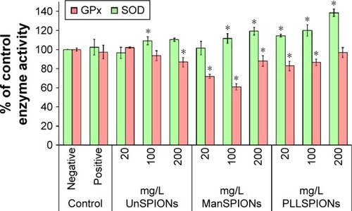
Figure 6 Effect of SPIONs on mitochondrial membrane potential measured by DiOC6 staining in neural stem cells after 4 hours of exposure.
Notes: The data, expressed as the mean of three independent experiments conducted in five replicates, were calculated as percentages of the values measured in negative controls (cells in nanoparticle-free exposure media). Positive controls are cells treated with 100 μM of hydrogen peroxide. Error bars represent standard deviation. All treatments were significantly different from negative controls at P<0.05.
Abbreviations: SPIONs, superparamagnetic iron oxide nanoparticles; UnSPIONs, uncoated SPIONs; ManSPIONs, D-mannose-coated SPIONs; PLLSPIONs, poly-L-lysine-coated SPIONs.
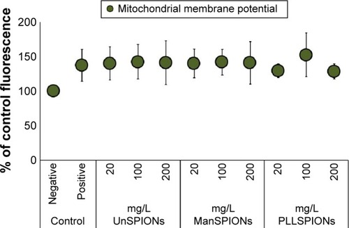
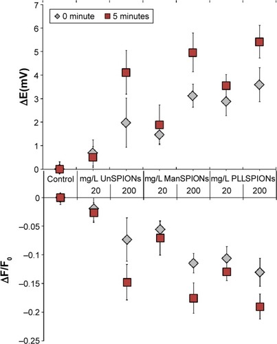
Figure 7 Effect of SPIONs on cell-membrane potential during 5 minutes of neural stem cell exposure.
Notes: The data, obtained as the mean of three independent experiments conducted in five replicates, were calculated as the difference between fluorescence intensities of treated and control (untreated) cells (ΔF/F0) divided by the initial fluorescence intensity (F0), and as changes in membrane potential (ΔE) in mV. Error bars represent standard deviation. All treatments were significantly different from controls at P<0.05.
Abbreviations: SPIONs, superparamagnetic iron oxide nanoparticles; UnSPIONs, uncoated SPIONs; ManSPIONs, D-mannose-coated SPIONs; PLLSPIONs, poly-L-lysine-coated SPIONs.