
Figures & data
Table 1 The composition of NPs for preliminary studies
Table 2 The composition of main NP formulations
Table 3 PS, PI, and ZP of NPs
Figure 1 TEM photographs of the NP formulations.
Notes: (A) N1 (the scale bar for image represents 500 nm), (B) N13 (the scale bar for image represents 5,000 nm), (C) N16 (the scale bar for image represents 20,000 nm), and (D) N19 (the scale bar for image represents 100 nm).
Abbreviations: TEM, transmission electron microscopy; NP, nanoparticle.
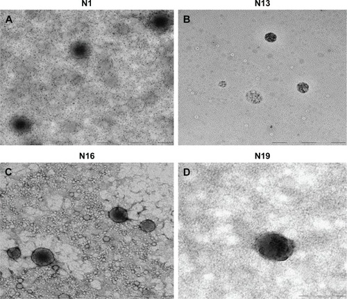
Figure 2 (A) Estimation of the ZP of the CSH-coated NPs during incubation in 0.1% aqueous mucin dispersion (B) estimation of the interaction between CSH-coated NPs and mucin dispersion by turbidimetric assay.
Abbreviations: ZP, zeta potential; NPs, nanoparticles; CSH, chitosan.
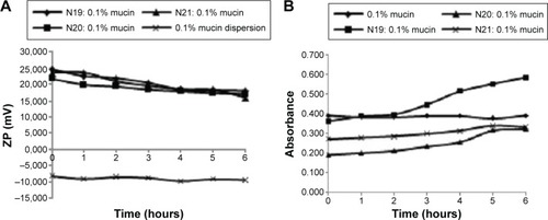
Table 4 EE and DL capacity of the NPs
Figure 3 (A) in vitro release of noncoated NPs and (B) in vitro release of CSH-coated NPs.
Abbreviations: FLZ, fluconazole; CSH, chitosan; NPs, nanoparticles.
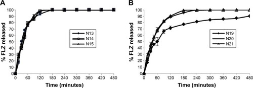
Table 5 Release parameters of FLZ from NPs
Figure 4 Cytotoxic effect of selected NPs evaluated by MTT assay.
Note: Bars represent “mean ± standard deviation” values from five individual experiments. *P<0.0005, group compared to control group (taken as 100%). Triton X-100 used as control for cytotoxic activity.
Abbreviations: NPs, nanoparticles; MTT, 3-(4,5-dimethylthiazol-2-yl)-2,5-diphenyltetrazolium bromide.
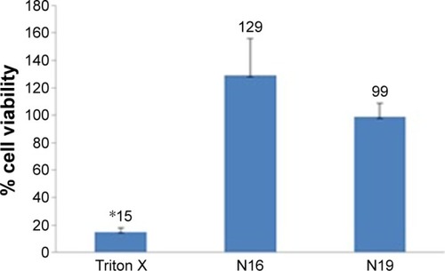
Figure 5 Zones caused by the formulations on G-MB-MHA medium (A) N16, (B) N19.
Abbreviation: G-MB-MHA, glucose- and methylene blue-added Mueller-Hinton Agar.
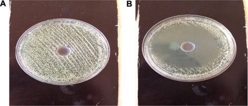
Figure 6 (A) Fungicidal effect of N19-related in vitro release study; (B) Time-dependent fungicidal effects of N19.
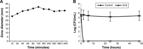
Figure 7 Images of histological examination.
Notes: (A) Palatal mucosa of a cured animal (1: epithelium, 2: lamina propria) (HE ×10). (B) Palatal mucosa of an animal with ongoing infection (1: epithelium, 2: lamina propria) (HE ×10). (C) Tongue mucosa of a cured animal (HE ×10) (1: epithelium, 2: lamina propria, 3: muscle, [→] papillae of the tongue). (D) Tongue mucosa of an animal with ongoing infection (HE ×10) (1: epithelium, 2: lamina propria, 3: muscle, [→] papillae of the tongue). (E) Buccal mucosa of a cured animal (HE ×10) (1: epithelium, 2: lamina propria, 3: muscle). (F) Buccal mucosa of an animal with ongoing infection (HE ×10) (1: epithelium, 2: lamina propria, 3: muscle, [→] degradation in the stratified epithelium).
Abbreviation: HE, hematoxylin–eosin.
![Figure 7 Images of histological examination.Notes: (A) Palatal mucosa of a cured animal (1: epithelium, 2: lamina propria) (HE ×10). (B) Palatal mucosa of an animal with ongoing infection (1: epithelium, 2: lamina propria) (HE ×10). (C) Tongue mucosa of a cured animal (HE ×10) (1: epithelium, 2: lamina propria, 3: muscle, [→] papillae of the tongue). (D) Tongue mucosa of an animal with ongoing infection (HE ×10) (1: epithelium, 2: lamina propria, 3: muscle, [→] papillae of the tongue). (E) Buccal mucosa of a cured animal (HE ×10) (1: epithelium, 2: lamina propria, 3: muscle). (F) Buccal mucosa of an animal with ongoing infection (HE ×10) (1: epithelium, 2: lamina propria, 3: muscle, [→] degradation in the stratified epithelium).Abbreviation: HE, hematoxylin–eosin.](/cms/asset/05cf8d4a-27f2-4474-b971-a1ac32aac4cc/dijn_a_103762_f0007_c.jpg)