Figures & data
Figure 1 RF-induced hyperthermia: experimental design.
Abbreviations: Dex-LSMO, dextran-coated lanthanum strontium manganese oxide; RF, radiofrequency; HSP, heat shock protein.
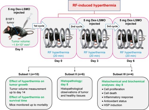
Figure 2 Qualitative assessment of Dex-LSMO nanoparticles distribution by magnetic resonance imaging.
Notes: Photographs (panel A) and processed ex vivo MR images (panel B) of representative tissues at different time points after intra-tumoral injection of Dex-LSMO nanoparticles. The gradient color bar on right indicates the signal level emanated by nanoparticles (lighter: lower concentration of nanoparticles; darker: higher concentration of nanoparticles).
Abbreviations: MR, magnetic resonance; Dex-LSMO, dextran-coated lanthanum strontium manganese oxide.
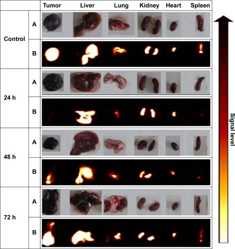
Figure 3 Quantitative assessment of Dex-LSMO nanoparticles distribution by atomic absorption spectroscopy.
Notes: Percent concentration of strontium (Sr conc) estimated at different time points (A) in tumor and (B) in different healthy tissues. *P<0.05 as analyzed by two-way ANOVA. Data are presented as mean ± SEM.
Abbreviations: ANOVA, analysis of variance; SEM, Standard error of the mean; Dex-LS MO, dextran-coated lanthanum strontium manganese oxide.
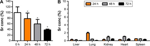
Figure 4 Effect of RF induced Dex-LSMO nanoparticles-mediated hyperthermia treatment on tumor growth and survival time.
Notes: (A) Photograph of melanoma-bearing mice in control and Dex-LSMO + RF-treated groups on Day 10. (B) Tumor volume of mice after treatment with Dex-LSMO-mediated hyperthermia as compared to control groups. *P<0.05, **P<0.01, and ***P<0.001 as analyzed by one-way ANOVA followed by Dunnett’s multiple comparison test. (C) Tumor inhibition percentage in different groups of mice on Day 14. (D) Kaplan–Meier survival curves of control and Dex-LSMO + RF-treated groups of mice. ***P<0.001 as analyzed by log rank test. Data are presented as mean ± SEM.
Abbreviations: Dex-LSMO, dextran-coated lanthanum strontium manganese oxide; RF, radiofrequency; ANOVA, analysis of variance; SEM, Standard error of the mean.
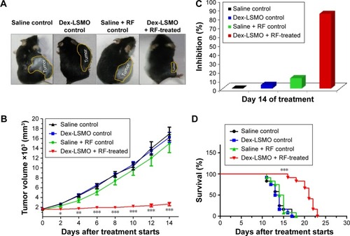
Figure 5 Effect of RF induced Dex-LSMO nanoparticles-mediated hyperthermia treatment on tumor histology.
Notes: Photomicrographs of HE-stained (A) tumor sections and (B) liver sections from untreated controls and Dex-LSMO + RF-treated mice on Day 8 of treatment. Asterisks show necrotic area.
Abbreviations: HE, hematoxylin and eosin; Dex-LSMO, dextran-coated lanthanum strontium manganese oxide; RF, radiofrequency.
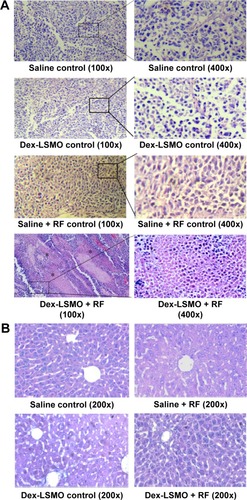
Figure 6 Effect of RF induced Dex-LSMO nanoparticles-mediated hyperthermia treatment on tumor proliferation.
Notes: Representative photomicrographs of Ki-67-stained tumor sections of saline control and Dex-LS MO + RF-treated mice.
Abbreviations: Dex-LSMO, dextran-coated lanthanum strontium manganese oxide; RF, radiofrequency.
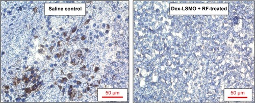
Figure 7 Assessment of apoptotic cell death after hyperthermia treatment.
Notes: (A) Representative images of TUNEL-positive apoptotic cells in tumor and liver sections of saline control and Dex-LSMO + RF-treated mice. (B) Agarose gel electrophoresis of DNA isolated from tumor of control and Dex-LSMO + RF-treated mice. (C) Caspase activity in tumor and liver tissues of saline control and Dex-LSMO + RF-treated mice. *P<0.05 as analyzed by unpaired Student’s t-test.
Abbreviations: TUNEL, terminal deoxynucleotidyl transferase-dUTP nick end labeling; Dex-LSMO, dextran-coated lanthanum strontium manganese oxide; RF, radio-frequency.
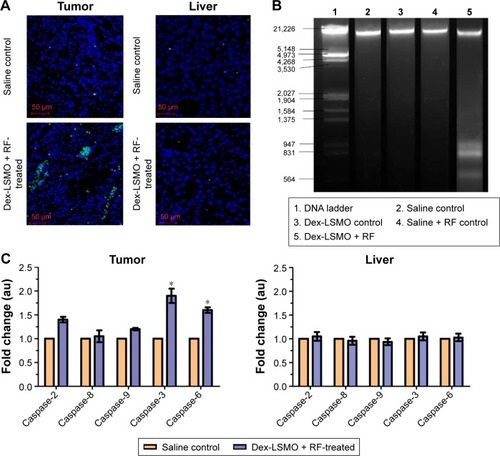
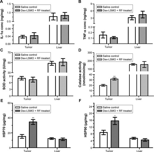
Figure 8 Effect of RF induced Dex-LSMO nanoparticles-mediated hyperthermia treatment on biochemical parameters.
Notes: Levels of (A) IL-1α and (B) TNF-α in tumor and liver of saline control and Dex-LSMO + RF-treated mice. (C) SOD and (D) catalase activity in tumor and liver of saline control and Dex-LSMO + RF-treated mice. Levels of (E) HSP70 and (F) HSP90 in tumor and liver of saline control and Dex-LSMO + RF-treated mice. *P<0.05 and **P<0.01 as analyzed by unpaired Student’s t-test.
Abbreviations: IL-1α, interleukin-1α; TNF-α, tumor necrosis factor-α; Dex-LSMO, dextran-coated lanthanum strontium manganese oxide; RF, radiofrequency; SOD, superoxide dismutase; HSP, heat shock protein.