Figures & data
Figure 1 a) TEM of GGS-NPs. Scale bar = 40 nm. b) Extinction spectrum of the GGS-NPs. c) GGS-NPs displayed a quadratic dependence of luminescence intensity on excitation power when exposed to an 800 nm pulsed laser.
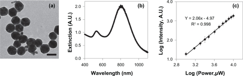
Figure 2 a) Two-photon induced photoluminescence images of SK-BR-3 cells exposed to 1 mW with the pulsed laser tuned to 800 nm. b) Brightfield images of SK-BR-3 cells in the same field-of-view as the luminescence images. c) Overlay of images (a) and (b), showing that luminescence was confined to cells targeted with anti-HER2 gold-gold sulfide nanoparticles. Scale bar = 100 μm.
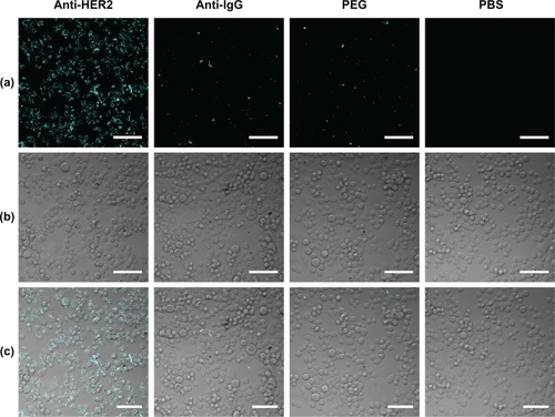
Figure 3 Calcein AM staining indicated that cancerous cells remained viable (evidenced by green fluorescent signal) when exposed to 1 mW laser power, regardless of nanoparticle presence. At 50 mW laser output a red fluorescent ethidium homodimer-1 signal indicative of membrane damage was observed in cells exposed to anti-HER2 functionalized GGS-NPs only where the laser was applied. Laser exposure alone was harmless to cells, as was laser exposure combined with nonspecifically targeted nanoparticles. Scale bar = 250 μm.
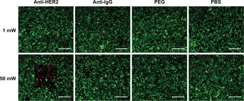
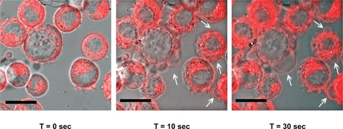
Figure 4 Time-lapse photography of SK-BR-3 cells exposed to anti-HER2 functionalized GGS-NPs and 50 mW laser power. The fluorescent red DiI membrane stain indicates regions of membrane blebbing generated by localized hyperthermia, with examples depicted by white arrows. Scale bar = 20 μm.