Figures & data
Figure 1 Characterization of G/SWCNT hybrids.
Notes: (A) TEM images of G/SWCNT hybrids showing the fibers and particles assembling together. (B) Raman spectrum of G/SWCNT hybrids.
Abbreviations: G/SWCNT, graphene/single-walled carbon nanotube; TEM, transmission electron microscopy.
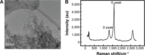
Figure 2 The viability and proliferation of rMSCs after they were treated with different concentrations of carbon nanomaterials.
Notes: (A) Cell viability of rMSCs incubated for 24 hours with different concentrations of G/SWCNT hybrids, G, and SWCNTs. (B) The proliferation of rMSCs after they were treated with different concentrations of G/SWCNT hybrids for 1, 3, and 7 days. Data represent mean ± SD for n=5. *P<0.05 (compared to control) or #P<0.05 (compared to the corresponding concentration of the G/SWCNT hybrids).
Abbreviations: G, graphene; G/SWCNT, graphene/single-walled carbon nanotube; NaF, sodium fluoride; rMSCs, rat mesenchymal stem cells; SD, standard deviation; SWCNTs, single-walled carbon nanotubes.
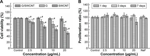
Figure 3 Immunofluorescence images of cytoskeletal organization of rMSCs stained with phalloidin for F-actin (red) and with PI for nuclei (blue).
Notes: (A) Cells incubated with culture medium were used as the control group. (B) Cells were pretreated with cytochalasin D for 30 minutes and then incubated (C) without and (D) with G/SWCNT hybrids at a concentration of 10 pg/mL for 24 hours. Scale bar =50 µm.
Abbreviations: G/SWCNT, graphene/single-walled carbon nanotube; PI, propidium iodide; rMSCs, rat mesenchymal stein cells.
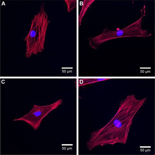
Figure 4 ALP activity of rMSCs treated with different concentrations of G/SWCNT hybrids.
Notes: Cells treated with G/SWCNT hybrids had higher ALP activity, indicating increased cellular differentiation. Data represent mean ± SD for n=4. *P<0.05 (compared to control).
Abbreviations: ALP, alkaline phosphatase; G/SWCNT, graphene/single-walled carbon nanotube; NaF, sodium fluoride; rMSCs, rat mesenchymal stem cells; SD, standard deviation.
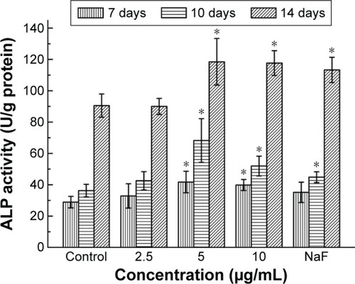
Figure 5 Effect of different concentrations of G/SWCNT hybrids on the mineralized bone nodule of rMSCs.
Notes: Representative images showing ARS of rMSCs treated (A) without and (B) with G/SWCNT hybrids after 14 days of differentiation. (C) NaF treatment was used as a positive control. (D) Cells incubated with the culture medium were used as the undifferentiation control. Scale bar =100 µm. (E) Mineralization was quantitated by measuring the ARS content of stained calcium deposits. The extracted ARS content confirmed that G/SWCNT hybrids increased extracellular calcium deposition of cells. Data represent the mean ± SD with n=4 for each bar. *P<0.05 (compared to control).
Abbreviations: ARS, alizarin red staining; G/SWCNT, graphene/single-walled carbon nanotube; NaF, sodium fluoride; rMSCs, rat mesenchymal stem cells; SD, standard deviation.
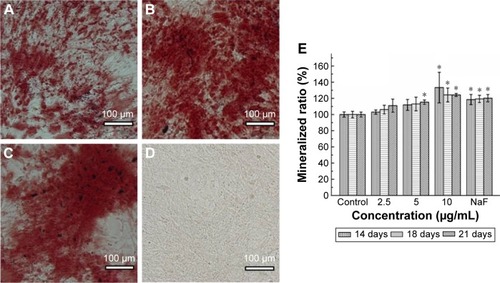
Figure 6 Evaluation of adipogenic differentiation of rMSCs after they were treated with G/SWCNT hybrids.
Notes: Representative images showing oil red O staining of adipocytes treated (A) without and (B) with G/SWCNT hybrids after 21 days of differentiation. (C) NaF treatment was used as a positive control. (D) Cells incubated with culture medium were used as the undifferentiation control. Scale bar =100 µm. (E) Adipocytes were quantitated by measuring oil red O from the stained cytoplasmic lipid accumulation. The results demonstrated G/SWCNT hybrids inhibited adipogenic differentiation of rMSCs. Data represent the mean ± SD with n=4 for each bar. *P<0.05 (compared to control).
Abbreviations: G/SWCNT, graphene/single-walled carbon nanotube; NaF, sodium fluoride; rMSCs, rat mesenchymal stem cells; SD, standard deviation.
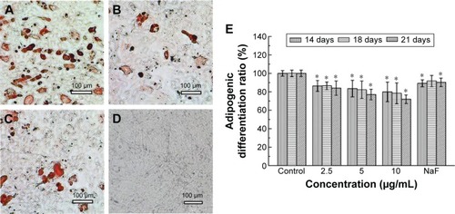
Figure 7 Western blot analysis for the expression levels of osteogenic and adipogenic differentiation specific genes in rMSCs treated with G/SWCNT hybrids after differentiation.
Notes: NaF-treated group served as positive control. After treatment with G/SWCNT hybrids, the expression levels of osteogenic genes, including Runx2, OCN, and OPN, were upregulated, while the adipose-specific gene, PPARγ2, was downregulated.
Abbreviations: C, control; GAPDH, glyceraldehyde-3-phosphate dehydrogenase; G/SWCNT, graphene/single-walled carbon nanotube; NaF, sodium fluoride; OCN, osteocalcin; OPN, osteopontin; PPARγ2, peroxisome proliferator-activated receptor-γ2; rMSCs, rat mesenchymal stem cells; Runx2, runt-related transcription factor 2.
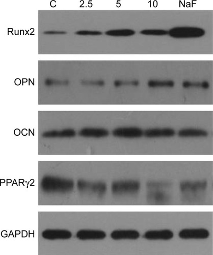
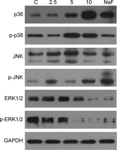
Figure 8 Western blot analysis for the expression levels of MAPK signaling pathway genes in rMSCs treated with G/SWCNT hybrids after differentiation.
Notes: The expression level of p38 was upregulated and ERK1/2 was downregulated in rMSCs in the presence of G/SWCNT hybrids.
Abbreviations: C, control; ERK1/2, extracellular signal-regulated protein kinases; GAPDH, glyceraldehyde-3-phosphate dehydrogenase; G/SWCNT, graphene/single-walled carbon nanotube; MAPK, mitogen-activated protein kinase; p-, phospho-; p38, p38 MAP kinase; rMSCs, rat mesenchymal stein cells.