Figures & data
Figure 1 Composition of ginsenosides in GBE.
Abbreviation: GBE, ginseng berry extract.
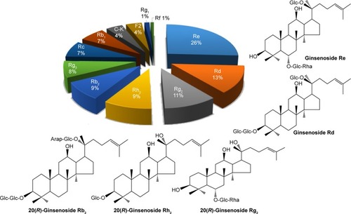
Figure 2 Phytochemical properties of Panax ginseng berry.
Abbreviations: GB, ginseng berry; UV, ultraviolet.
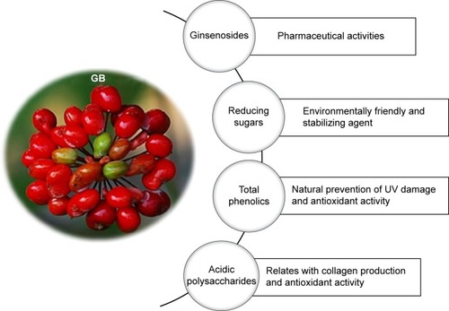
Figure 3 Reaction temperature and concentration of GBE have been successfully optimized to maximize the yield of nanoparticles.
Notes: Temperature-dependent evolution of UV–vis spectra of synthesized nanoparticles at a constant amount of GBE (3%, v/v) and 1 mM of gold and silver salts: (A) GBAuNPs and (B) GBAgNPs. Concentration-dependent evolution of UV–vis at 80°C of (C) GBAuNPs (D) GBAgNPs. Time-dependent evolution of UV–vis at 80°C, GBE 5% concentration of GBE and the respective photograph which shows the color change pattern during nanoparticle synthesis of (E) GBAuNPs and (F) GBAgNPs.
Abbreviations: GBAgNPs, silver nanoparticles from ginseng berry; GBAuNPs, gold nanoparticles from ginseng berry; GBE, ginseng berry extract; OD, optical density; UV, ultraviolet; vis, visible.
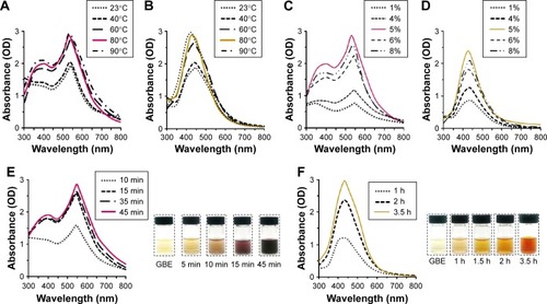
Table 1 Rate constants at different temperatures
Figure 4 FE-TEM imaging shows that the particles are spherical in shape.
Notes: (A) GBAuNPs with sizes between 5 and 10 nm and (B) GBAgNPs with sizes between 10 and 20 nm. EDX spectrum of (C) GBAuNPs confirmed the presence of characteristic peak of metallic gold at 2.2 keV and (D) GBAgNPs characteristic peak of silver at 3.3 keV. Elemental mapping analysis of (E) GBAuNPs and (F) GBAuNPs showed maximum distribution of gold (green) and silver (red) elements in the corresponding nanoparticles.
Abbreviations: EDX, energy-dispersive X-ray spectrometer; FE-TEM, field-emission transmission electron microscopy; GBAgNPs, silver nanoparticles from ginseng berry; GBAuNPs, gold nanoparticles from ginseng berry.
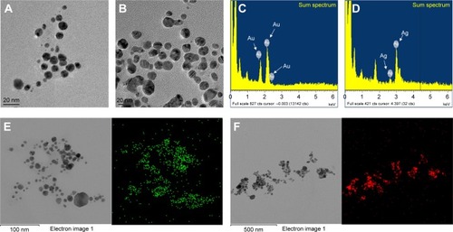
Figure 5 XRD pattern for the identification of crystallinity exhibited by the synthesized materials.
Notes: (A) GBAuNPs and (B) GBAgNPs. SAED pattern indicated that an FCC crystal structure of (C) single GBAuNPs is spherical and (D) single GBAgNPs is spherical.
Abbreviations: FCC, face-centered cubic; GBAgNPs, silver nanoparticles from ginseng berry; GBAuNPs, gold nanoparticles from ginseng berry; SAED, selected area electron diffraction; XRD, X-ray diffraction.
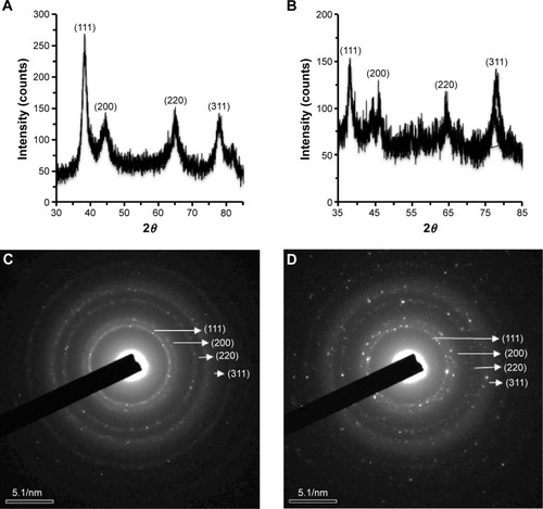
Figure 6 FTIR spectra of (A) GBE, (B) GBAgNPs and (C) GBAuNPs.
Notes: The peaks of polysaccharide and hydroxyl group of phenolic compound structures due to O–H stretching are shown between 3,500 and 3,000 cm−1. C–H stretching between 3,000 and 2,800 cm−1 corresponds to methyl and methylene group, and peaks between 1,600 and 1,400 cm−1 represent carbonyl group stretching vibration of flavonoids and ester bonds in polyphenolic compounds. Weak peaks between 1,500 and 1,032 cm−1 corresponds to aromatic C–C groups and C–O functional in tanning/tannic acid.
Abbreviations: FTIR, Fourier transform infrared; GBAgNPs, silver nanoparticles from ginseng berry; GBAuNPs, gold nanoparticles from ginseng berry; GBE, ginseng berry extract.
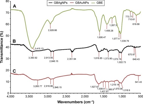
Figure 7 AuNPs and AgNPs synthesized from GBE exhibited free radical scavenging activity.
Notes: (A) Comparative DPPH radical scavenging activity of gallic acid, GBAgNPs, GBAuNPs, GBE, and gold and silver salts. GBAgNPs showed the highest free radical scavenging activity (93.8%) compared to GBAuNPs (81.5%) and GBE (81%). AgNPs have antimicrobial activity against Gram-negative (E. coli) and Gram-positive (S. aureus) bacterial strains. (B) Antibacterial activity of GBAgNPs of various concentrations (1, 15 μg/disk; 2, 30 μg/disk and 3, 45 μg/disk) of synthesized GBAgNPs using GBE. Neomycin was used as assay control.
Abbreviations: AgNPs, silver nanoparticles; AuNPs, gold nanoparticles; C, control; DPPH, 2,2-diphenyl-1-picrylhydrazyl; E. coli, Escherichia coli; GA, gallic acid; GBAgNPs, AgNPs from ginseng berry; GBAuNPs, AuNPs from ginseng berry; GBE, ginseng berry extract; S. aureus, Staphylococcus aureus.
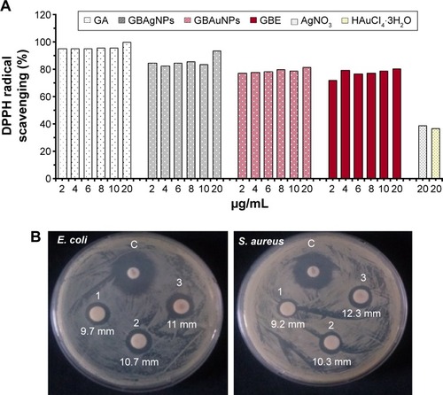
Table 2 Tyrosinase inhibitory activity using l-DOPA as substrate
Figure 8 Comparative study of the effect of GBAuNPs, GBE and HAuCl4·3H2O on cell viability and on the morphology of HDF and B16 cells.
Notes: Optical microscopy images (A) of HDF and B16 cell lines (40× magnification) at 72 h after treated with 1–100 μg/mL of GBAuNPs. Treated HDF and B16 cells with AuNPs (10 and 100 μg/mL) could be identified by dark dense clusters which are indicated with arrows; no cluster was found in control groups. Cytotoxicity effect of GBE and GBAuNPs on HDF (B) and B16BL6 (C) cell lines; GBAuNPs did not exhibit a significant cytotoxic effect on HDF and B16 cells. Cell viability was determined by MTT assay. Data are expressed as a percentage of sample-treated control and presented as mean ± SEM of three separate experiments. B16, murine melanoma B16BL6 cells.
Abbreviations: AuNPs, gold nanoparticles; GBAuNPs, AuNPs from ginseng berry; GBE, ginseng berry extract; HDF, human dermal fibroblast; SEM, standard error of the mean.
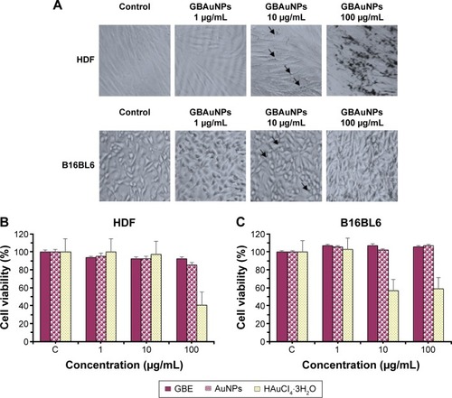
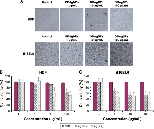
Figure 9 Comparative study of the effect of GBAgNPs, GBE and AgNO3 on cell viability and on the morphology of HDF and B16 cells.
Notes: Optical microscopy images (A) of HDF and B16 cell lines (40× magnification) at 72 h after treated with 1–100 μg/mL of GBAgNPs. Treated HDF and B16 cells with silver (10 μg/mL) could be identified by dark dense clusters which are indicated with arrows; no cluster was found in control groups. Cytotoxicity effect of GBE, GBAgNPs, and silver salts on HDF (B) and B16 (C) cell lines; cell viability decreased with an increase in the concentration of GBAgNPs. Cell viability was determined by MTT assay. Data are expressed as a percentage of sample-treated control and presented as mean ± SEM of three separate experiments.
Abbreviations: B16, murine melanoma B16Bl6 cells; GBAgNPs, silver nanoparticles from ginseng berry; GBE, ginseng berry extract; HDF, human dermal fibroblast; MTT, 3-(4,5-dimethyl-thiazol-2yl)-2, 5-diphenyl tetrazolium bromide; SEM, standard error of the mean.