Figures & data
Figure 1 Synthesis of LHRH-PEG-DSPE and preparation of LHRH-MTX-SL.
Abbreviations: LHRH, luteinizing hormone-releasing hormone; PEG, polyethylene glycol; MTX, mitoxanthrone; SL, loaded liposomes.
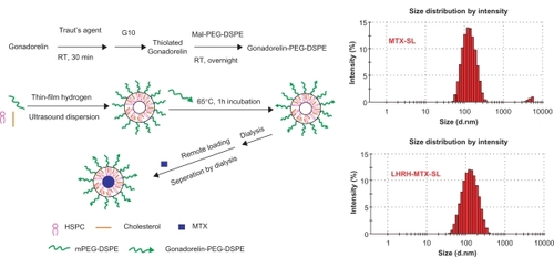
Table 1 Particle size, polydispersity index, zeta potential and gonadorelin:lipid ratio of liposomal formulations with or without mitoxantrone. The dynamic light scattering analysis was performed at 25°C and at a scattering angle of 90°. The obtained particle suspension was diluted appropriately in phosphate-buffered saline before measurement
Figure 2 Release profiles of mitoxantrone from MTX-SL and LHRH-MTX-SL in phosphate-buffered saline A or isotonic glucose containing 10% human plasma B Release of mitoxantrone from different liposome preparations was evaluated by a dialysis method, followed by high-performance liquid chromatography analysis for determination of mitoxantrone. Results are expressed as means ± standard error of measurement of three independent measurements.
Abbreviations: LHRH, luteinizing hormone-releasing hormone; MTX, mitoxanthrone; SL, loaded liposomes.
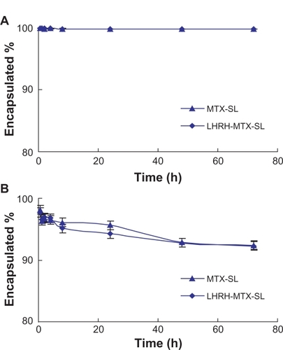
Figure 3 Uptake of different liposomes encapsulating mitoxantrone in various cell lines by confocal laser scanning fluorescence microscopy. Various cell lines were treated with either LHRH-MTX-SL or MTX-SL for four hours at 37°C. A LHRH-MTX-SL in LHRH receptor high-expressing MCF-7 cells; B MTX-SL in LHRH receptor high-expressing MCF-7 cells; C MCF-7 cells treated with drug-free medium used as a control; D LHRH-MTX-SL in LHRH receptor low-expressing SK-OV-3 cells; E MTX-SL in LHRH receptor low-expressing SK-OV-3 cells; F SK-OV-3 cells treated with drug-free medium used as a control.
Abbreviations: LHRH, luteinizing hormone-releasing hormone; MTX, mitoxanthrone; SL, loaded liposomes.
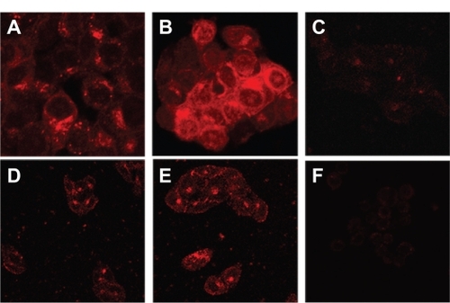
Figure 4 Viability of MCF-7 cells after treatment with mitoxantrone, mitoxantrone-containing liposomes, and empty liposomes for 24 hours A and 120 hours B The cells were exposed to serial concentrations of free mitoxantrone, MTX-SL, LHRH-MTX-SL at 37°C for 24 hours or 120 hours. The drug concentration of free mitoxantrone and MTX-SL used in this assay was 0.3, 3.0, 30, 300 ng/mL. Empty liposomes were used as a control. The antitumor effect was evaluated usings the MTT method. MCF-7 cell viability (as a percentage of control cells) was calculated as follows: Viability(%) = Atreated/Acontrol × 100%. Results are expressed as means ± standard error of measurement from six independent measurements.
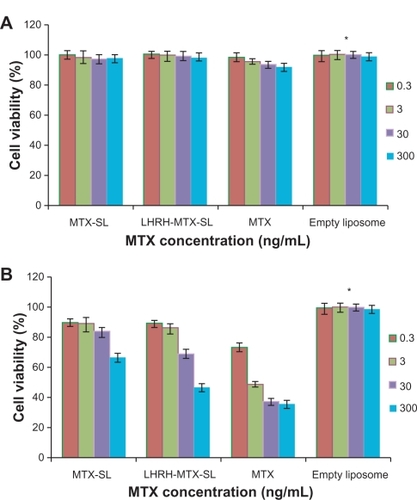
Table 2 Cytotoxicity data (IC50) for free mitoxantrone and mitoxantrone liposomal formulations (with or without LHRH) determined with MCF-7 cells (n = 3). Data are represented as percentage of control. Each assay was done in triplicate (mean ± standard error of measurement). The cells were exposed to serial concentrations of free mitoxantrone, MTX-SL, and LHRH-MTX-SL at 37°C for 120 hours. The cytotoxicity was evaluated using the MTT method. MCF-7 cell viability (as a percentage of control cells) was calculated as follows: Viability (%) = Atreated/Acontrol × 100%. IC50 was defined as the concentration of drug where cell growth and/or viability was 50% of that observed in control (drug-free) cultures in the MTT assay