Figures & data
Figure 1 Schematic of the preparation process of Mt-BH-LPs.
Abbreviations: Mt-BH-LPs, montmorillonite–betaxolol hydrochloride liposomes; BH, betaxolol hydrochloride; Mt, montmorillonite.
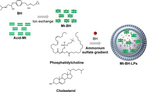
Figure 2 Schematic diagram of the in vitro tear-turnover apparatus.
Notes: The model incorporated an insert containing cultured corneal tissue as a turnover chamber; the external basal side of the insert was sealed to avoid a downward diffusion of material. The system was temperature-controlled at human tear-film temperature (34°C). Constant in- and outflow of simulated tears in the chamber were controlled by peristaltic pumps.
Abbreviations: STF, simulated tear fluid; iHCEC, immortalized human corneal epithelial cell.
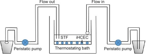
Figure 3 Time to dialysis equilibrium of Mt-BH-LPs
Abbreviation: Mt-BH-LPs, montmorillonite–betaxolol hydrochloride liposomes.

Figure 4 Particle-size distribution of (A) BH-LPs and (B) Mt-BH-LPs.
Note: The red line indicates cumulative distribution. The black line inidates the percentage of the particle size. f, differential intensity.
Abbreviation: Mt-BH-LPs, montmorillonite–betaxolol hydrochloride liposomes.
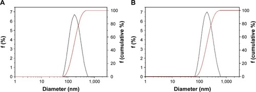
Figure 5 TEM images of (A, B) BH-LPs and (C, D) Mt-BH-LPs.
Note: (B and D) show magnification of red squares in (A and C), respectively.
Abbreviations: TEM, transmission electron microscopy; Mt-BH-LPs, montmorillonite–betaxolol hydrochloride liposomes.
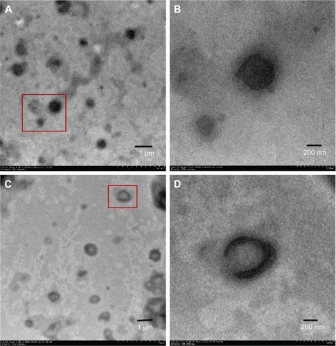
Figure 6 (A) XRD patterns of BH, acid-Mt, Mt-BH, acid-Mt + BH BH-LPs, and Mt-BH-LPs; (B) enlarged XRD patterns of acid-Mt and Mt-BH.
Abbreviations: XRD, X-ray diffraction; Mt-BH-LPs, montmorillonite–betaxolol hydrochloride liposomes; BH, betaxolol hydrochloride; Mt, montmorillonite.
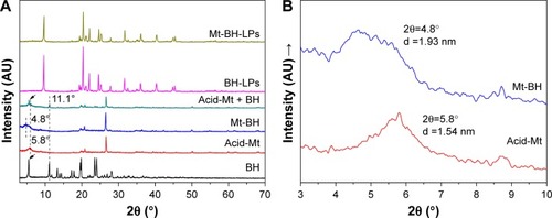
Figure 7 (A) TGA and (B) DSC pattern of BH, acid-Mt, Mt-BH, BH-LPs, and Mt-BH-LPs.
Abbreviations: TGA, thermogravimetric analysis; DSC, differential scanning calorimetry; Mt-BH-LPs, montmorillonite–betaxolol hydrochloride liposomes; BH, betaxolol hydrochloride; Mt, montmorillonite.
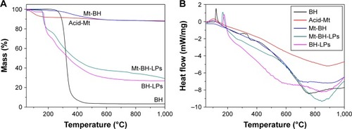
Figure 8 FTIR spectra of acid-Mt, BH, Mt-BH, and Mt-BH-LPs.
Abbreviations: FTIR, Fourier-transform infrared; Mt-BH-LPs, montmorillonite–betaxolol hydrochloride liposomes; BH, betaxolol hydrochloride; Mt, montmorillonite.
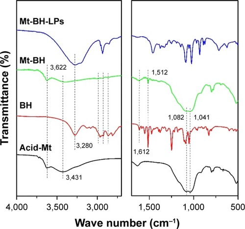
Figure 9 In vitro release curves of BH solution, BH-LPs, and Mt-BH-LPs.
Abbreviations: Mt-BH-LPs, montmorillonite–betaxolol hydrochloride liposomes; BH, betaxolol hydrochloride.
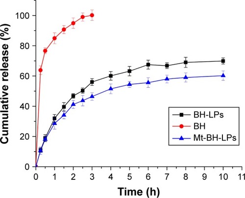
Figure 10 Cell viability of (A) Betoptic, (B) BH solution, (C) blank LPs, and (D) Mt-BH-LPs at different exposure times and amounts.
Abbreviations: Mt-BH-LPs, montmorillonite–betaxolol hydrochloride liposomes; BH, betaxolol hydrochloride.
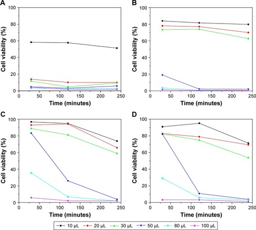
Figure 11 Viability of iHCECs with differing amounts of Betoptic, BH solution, blank LPs, and Mt-BH-LPs exposure for 2 hours.
Abbreviations: iHCECs, immortalized human corneal epithelial cells; Mt-BH-LPs, montmorillonite–betaxolol hydrochloride liposomes; BH, betaxolol hydrochloride.
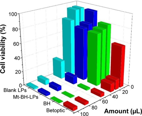
Table 1 Draize test scores
Figure 12 Cornea histopathology by microscopy.
Notes: (A) Normal cornea, (B) treated with saline, (C) treated with Betoptic, (D) treated with BH solution, and (E) treated with Mt-BH-LPs.
Abbreviations: Mt-BH-LPs, montmorillonite–betaxolol hydrochloride liposomes; BH, betaxolol hydrochloride.
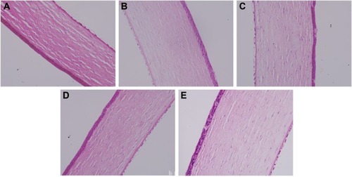
Figure 13 Conjunctival histopathology by microscopy.
Notes: (A) Normal cornea, (B) treated with saline, (C) treated with Betoptic, (D) treated with BH solution, and (E) treated with Mt-BH-LPs.
Abbreviations: Mt-BH-LPs, montmorillonite–betaxolol hydrochloride liposomes; BH, betaxolol hydrochloride.
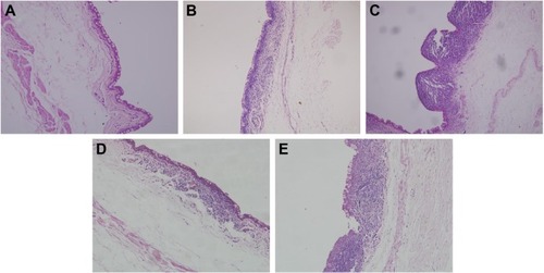
Figure 14 (A) Hemorrhage situation and (B) trypan blue absorption of CAM.
Abbreviations: NS, normal saline; Mt-BH-LPs, montmorillonite–betaxolol hydrochloride liposomes; BH, betaxolol hydrochloride; CAM, chorioallantoic membrane.
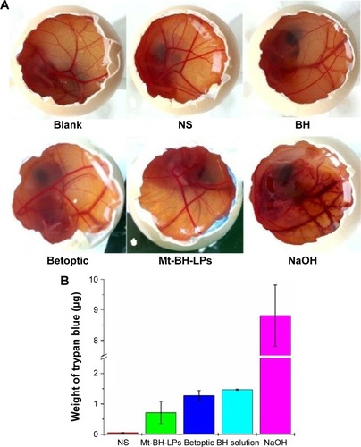
Figure 15 Frozen section of stratified immortalized human cornea epithelial cells.
Note: Fluorescence microscope image (magnification 400×).
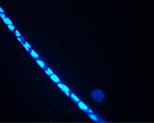
Figure 16 Concentration–time curve in the cornea/tear-film compartment after topical application of BH solution and Mt-BH-LPs.
Abbreviations: Mt-BH-LPs, montmorillonite–betaxolol hydrochloride liposomes; BH, betaxolol hydrochloride.
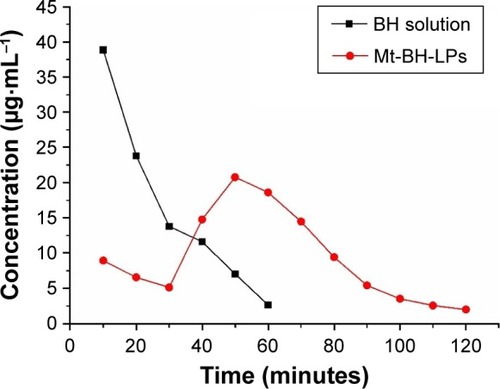
Table 2 Pharmacokinetics parameters of BH in tears after topical administration in rabbits
Figure 17 Mean tear-fluid concentration–time curve after topical application of BH solution and Mt-BH-LPs in rabbit eyes.
Abbreviations: Mt-BH-LPs, montmorillonite–betaxolol hydrochloride liposomes; BH, betaxolol hydrochloride.
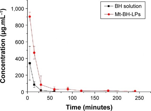
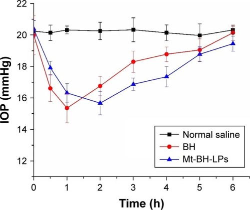
Figure 18 Change in IOP for rabbits with saline, BH solution, and Mt-BH-LPs.
Abbreviations: IOP, intraocular pressure; Mt-BH-LPs, montmorillonite–betaxolol hydrochloride liposomes; BH, betaxolol hydrochloride.