Figures & data
Figure 1 Diagrams of three types of organic–inorganic hybrid nanocomposites based on polymer, LDHs and drug.
Notes: (A) Organic-coated inorganic hybrid LDH nanoparticles. (B) Organic-intercalated inorganic hybrid LDH nanoparticles. (C) Organic–inorganic hybrid LDH nanosheets.
Abbreviation: LDH, layered double hydroxide.
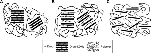
Table 1 Characterization of different types of CG-VV-LDH nanosheets
Figure 2 Cumulative release–time profiles of PRN and CG-VV-PRN-LDH nanosheets.
Note: Each value represents mean ± SD (n=3).
Abbreviations: PRN, pirenoxine sodium; CG-VV, chitosan–glutathione–valine–valine; LDH, layered double hydroxide; SD, standard deviation.
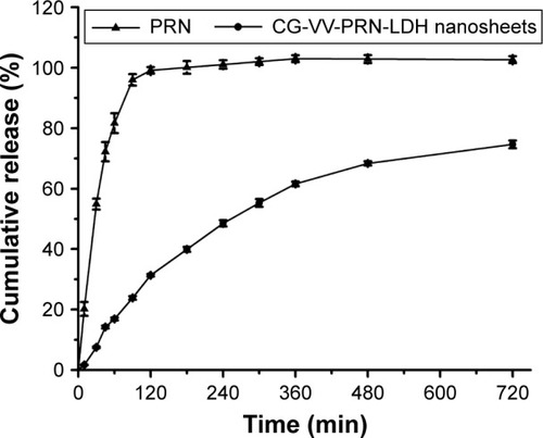
Table 2 Stability of CG-VV-PRN-LDH nanosheet eye drops
Table 3 Pharmacokinetic parameters of commercial PRN eye drops and CG-VV-PRN-LDH nanosheet eye drops
Figure 3 The concentration–time curve of PRN in tear samples of commercial PRN eye drops and CG-VV-PRN-LDH nanosheet eye drops.
Note: Each value represents mean ± SD (n=6).
Abbreviations: PRN, pirenoxine sodium; CG-VV, chitosan–glutathione–valine– valine; LDH, layered double hydroxide; SD, standard deviation.
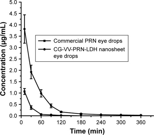
Figure 4 In vitro corneal permeation profiles of commercial PRN eye drops and CG-VV-PRN-LDH nanosheet eye drops in GBR solution.
Note: Each value represents mean ± SD (n=6).
Abbreviations: PRN, pirenoxine sodium; CG-VV, chitosan–glutathione–valine– valine; LDH, layered double hydroxide; GBR, glutathione-sodium bicarbonate Ringer; SD, standard deviation.
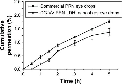
Figure 5 In vitro cytotoxicity of LDH nanoparticles, LDH nanosheets, CG-VV-LDH nanoparticles and CG-VV-LDH nanosheets against (A) HCEpiC, (B) ARPE-19 cells; (C) in vitro cytotoxicity of PRN-LDH nanoparticles, CG-VV-PRN-LDH nanoparticles and CG-VV-PRN-LDH nanosheets against HCEpiC after 12 h incubation, respectively; cell viability was determined by MTT assay.
Note: Each value represents mean ± SD (n=4).
Abbreviations: LDH, layered double hydroxide; CG-VV, chitosan–glutathione–valine–valine; HCEpiC, human corneal epithelial primary cells; ARPE-19, retinal pigment epithelial; PRN, pirenoxine sodium; MTT, 3-(4,5-dimethyl-2-thiazolyl)-2,5-diphenyl-2-H-tetrazolium bromide; SD, standard deviation.
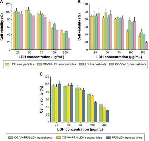
Figure 6 Fluorescence microscopy images of (A) HCEpiC and (B) ARPE-19 cells incubated with the following: a, FITC-LDH nanoparticles; b, CG-VV-FITC-LDH nanoparticles; c, CG-VV-FITC-LDH nanosheets; d and e, CG-VV-FITC solution (0.008% and 0.00038% [w/v]); f, free FITC at 37°C for 1, 2 and 4 h, respectively.
Note: Scale bar =25 μm.
Abbreviations: HCEpiC, human corneal epithelial primary cells; ARPE-19, retinal pigment epithelial; FITC, fluorescein isothiocyanate; LDH, layered double hydroxide; CG-VV, chitosan–glutathione–valine–valine.
![Figure 6 Fluorescence microscopy images of (A) HCEpiC and (B) ARPE-19 cells incubated with the following: a, FITC-LDH nanoparticles; b, CG-VV-FITC-LDH nanoparticles; c, CG-VV-FITC-LDH nanosheets; d and e, CG-VV-FITC solution (0.008% and 0.00038% [w/v]); f, free FITC at 37°C for 1, 2 and 4 h, respectively.Note: Scale bar =25 μm.Abbreviations: HCEpiC, human corneal epithelial primary cells; ARPE-19, retinal pigment epithelial; FITC, fluorescein isothiocyanate; LDH, layered double hydroxide; CG-VV, chitosan–glutathione–valine–valine.](/cms/asset/e33c4972-88e7-4e15-a2a6-1db5c6b65f7a/dijn_a_129311_f0006_c.jpg)
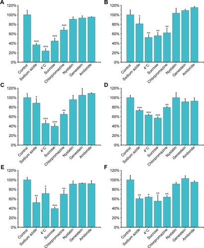
Figure 7 Flow cytometry measurement of intracellular uptake of (A) FITC-LDH nanoparticles, (B) CG-VV-FITC-LDH nanoparticles, (C) CG-VV-FITC-LDH nanosheets treated with specific endocytic inhibitors on HCEpiC; flow cytometry measurement of intracellular uptake of (D) FITC-LDH nanoparticles, (E) CG-VV-FITC-LDH nanoparticles, (F) CG-VV-FITC-LDH nanosheets treated with specific endocytic inhibitors on ARPE-19 cells.
Notes: HCEpiC or ARPE-19 cells without treatment with specific endocytic inhibitors were taken as the control (*P<0.05, **P<0.01, ***P<0.001 vs control group, n=3).
Abbreviations: FITC, fluorescein isothiocyanate; LDH, layered double hydroxide; CG-VV, chitosan–glutathione–valine–valine; HCEpiC, human corneal epithelial primary cells; ARPE-19, retinal pigment epithelial.