Figures & data
Figure 1 pH-responsive ACPP-modified liposome and its tumor-targeted delivery strategy.
Notes: The siRNA-loaded liposomes are retained in the tumor site due to the EPR effect. The acidic pHe in the tumor microenvironment splits ACPP at the hydrazone site and detaches the shielding domain from the CPP section, thereby recovering the potency of CPP for enhanced cellular internalization and endosome/lysosome escape.
Abbreviations: ACPP, activatable cell-penetrating peptide; CPP, cell-penetrating peptide; DSPE, distearoyl phosphatidylethanolamine; EPR, enhanced permeability and retention; PEG, polyethylene glycol; pHe, extracellular pH; siRNA, small interfering RNA.
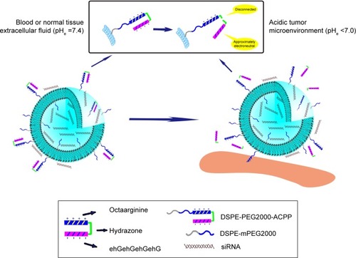
Figure 2 Procedure for the synthesis of ACPP.
Abbreviations: ACPP, activatable cell-penetrating peptide; Arg, arginine; CPP, cell-penetrating peptide; Cys, cysteine; Glu, glutamic acid; Gly, glycine; His, histidine; MPH, 3-maleimidopropionic acid hydrazide-trifluoroacetic acid; NAPM, N-(4-acetylphenyl)maleimide; N-M, NAPM–MPH; NP, cysteine–glycine–[(d-glutamic acid)–(d-histidine)–(d-glycine)]4.
![Figure 2 Procedure for the synthesis of ACPP.Abbreviations: ACPP, activatable cell-penetrating peptide; Arg, arginine; CPP, cell-penetrating peptide; Cys, cysteine; Glu, glutamic acid; Gly, glycine; His, histidine; MPH, 3-maleimidopropionic acid hydrazide-trifluoroacetic acid; NAPM, N-(4-acetylphenyl)maleimide; N-M, NAPM–MPH; NP, cysteine–glycine–[(d-glutamic acid)–(d-histidine)–(d-glycine)]4.](/cms/asset/5b688285-46de-4231-a171-c4101186498c/dijn_a_129574_f0002_b.jpg)
Figure 3 Identification of ACPP and its intermediate products.
Notes: (A) Citation1H-NMR spectrum of N-M in DMSO-d6. The positive ion electrospray ionization mass spectrum of (B) N-M and (C) NP-N-M. (D) MALDI-TOF mass spectrum of ACPP.
Abbreviations: ACPP, activatable cell-penetrating peptide; DMSO-d6, deuterated dimethyl sulfoxide; Citation1H-NMR, Citation1H nuclear magnetic resonance; MALDI-TOF, matrix-assisted laser desorption/ionization time-of-flight; MPH, 3-maleimidopropionic acid hydrazide-trifluoroacetic acid; NAPM, N-(4-acetylphenyl)maleimide; N-M, NAPM–MPH; NP, cysteine–glycine–[(d-glutamic acid)–(d-histidine)–(d-glycine)]4.
![Figure 3 Identification of ACPP and its intermediate products.Notes: (A) Citation1H-NMR spectrum of N-M in DMSO-d6. The positive ion electrospray ionization mass spectrum of (B) N-M and (C) NP-N-M. (D) MALDI-TOF mass spectrum of ACPP.Abbreviations: ACPP, activatable cell-penetrating peptide; DMSO-d6, deuterated dimethyl sulfoxide; Citation1H-NMR, Citation1H nuclear magnetic resonance; MALDI-TOF, matrix-assisted laser desorption/ionization time-of-flight; MPH, 3-maleimidopropionic acid hydrazide-trifluoroacetic acid; NAPM, N-(4-acetylphenyl)maleimide; N-M, NAPM–MPH; NP, cysteine–glycine–[(d-glutamic acid)–(d-histidine)–(d-glycine)]4.](/cms/asset/4dec421f-dc7b-4dcf-824a-f84e6ea98948/dijn_a_129574_f0003_c.jpg)
Figure 4 pH-sensitive profiles of ACPP.
Note: The responses of ACPP were plotted against incubation time at 37°C.
Abbreviation: ACPP, activatable cell-penetrating peptide.
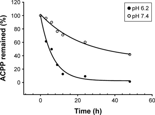
Figure 5 Synthesis route and identification of DSPE-PEG2000-ACPP/DSPE-PEG2000-CPP.
Notes: (A) Synthesis route of DSPE-PEG2000-ACPP/DSPE-PEG2000-CPP. MALDI-TOF mass spectrum of (B) DSPE-PEG2000-ACPP and (C) DSPE-PEG2000-CPP.
Abbreviations: ACPP, activatable cell-penetrating peptide; CPP, cell-penetrating peptide; DSPE, distearoyl phosphatidylethanolamine; MALDI-TOF, matrix-assisted laser desorption/ionization time-of-flight; NHS, N-hydroxysuccinimide; PEG, polyethylene glycol; TEA, triethylamine.
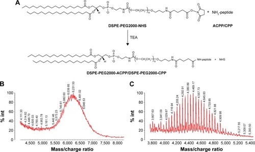
Table 1 Physicochemical properties of liposomes carrying siRNA
Figure 6 Micrographs of A-L.
Notes: (A) TEM image of A-L. AFM topographic images of A-L in (B) the adhesion channel and (C) the height sensor channel obtained by quantitative nanomechanical analysis.
Abbreviations: ACPP, activatable cell-penetrating peptide; AFM, atomic force microscopy; A-L, ACPP-modified liposomes; TEM, transmission electron microscopy.
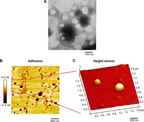
Figure 7 Flow cytometric measurement of liposomal FAM-siRNA uptake by MCF-7 cells.
Notes: MCF-7 cells were collected following 2 h of incubation in pH 6.2 or 7.4 medium containing various FAM-siRNA-loaded liposomes at 37°C. All liposomes used underwent 5 h of preincubation in serum-free medium at the pH corresponding to the subsequent exposure. The concentration of FAM-siRNA was 100 nM. ***P<0.001 (n=3).
Abbreviations: ACPP, activatable cell-penetrating peptide; A-L, ACPP-modified liposomes; C-L, CPP-modified liposomes; CPP, cell-penetrating peptide; N-L, nonmodified liposomes; ns, not significant; siRNA, small interfering RNA.
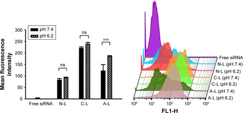
Figure 8 CLSM analysis of liposomal FAM-siRNA uptake by MCF-7 cells.
Notes: MCF-7 cells were incubated with pH 6.2 or 7.4 medium containing various FAM-siRNA-loaded liposomes at 37°C for 3.5 h. All liposomes were subject to the same pre-treatment as the liposomal formulations evaluated by flow cytometry. The FAM-siRNA concentration was 200 nM. Cell nuclei and F-actin were counterstained with Hoechst 33258 (blue) and rhodamine-phalloidin (red), respectively. FAM-siRNA fluorescence (green) was recorded.
Abbreviations: ACPP, activatable cell-penetrating peptide; A-L, ACPP-modified liposomes; C-L, CPP-modified liposomes; CLSM, confocal laser-scanning microscopy; CPP, cell-penetrating peptide; FAM-siRNA, FAM-labeled siRNA; N-L, nonmodified liposomes; siRNA, small interfering RNA.
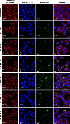
Figure 9 Intracellular trafficking and distribution of FAM-siRNA in (A) MCF-7 cells and (B) A549 cells.
Notes: The cells were treated with FAM-siRNA-encapsulated A-L at 37°C. The FAM-siRNA concentration was 200 nM. Cell nuclei, early endosomes, and late endosomes/lysosomes were counterstained with Hoechst 33258 (blue), CellLight Early Endosomes-RFP BacMam 2.0 (red), and LysoTracker Red (red), respectively. FAM-siRNA fluorescence (green) was recorded.
Abbreviations: ACPP, activatable cell-penetrating peptide; A-L, ACPP-modified liposomes; DIC, differential interference contrast; FAM-siRNA, FAM-labeled small interfering RNA.
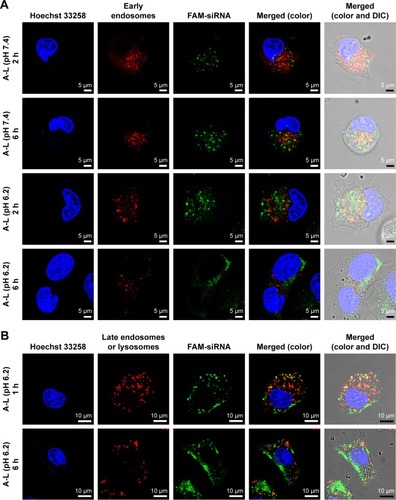
Figure 10 Cell apoptosis following exposure to different liposome formulations.
Notes: MCF-7 cells were individually treated with various formulations carrying siPLK-1 or siN.C. (100 nM) at 37°C for 6 h, followed by 72 h of routine culture. *P<0.05 (n=3). Early apoptotic cells are shown in the lower right quadrant, and late apoptotic cells are shown in the upper right quadrant.
Abbreviations: ACPP, activatable cell-penetrating peptide; A-L, ACPP-modified liposomes; C-L, CPP-modified liposomes; CPP, cell-penetrating peptide; mRNA, messenger RNA; N-L, nonmodified liposomes; ns, not significant; siN.C., negative control siRNA; siPLK-1, siRNA targeting polo-like kinase 1 mRNA.
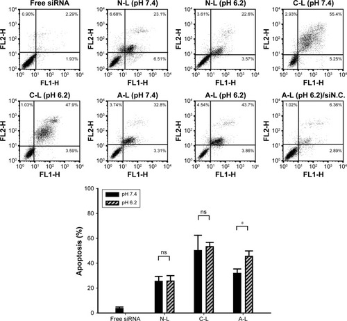
Figure 11 Analysis of PLK-1 and PLK-1 mRNA expression.
Notes: (A) The level of PLK-1 mRNA determined by qRT-PCR. (B) PLK-1 protein expression determined by Western blot analysis. MCF-7 cells were individually treated as described in the legend to for the cell apoptosis analysis, followed by (A) 48 h or (B) 72 h of routine culture. *P<0.05 (n=3).
Abbreviations: ACPP, activatable cell-penetrating peptide; A-L, ACPP-modified liposomes; C-L, CPP-modified liposomes; CPP, cell-penetrating peptide; mRNA, messenger RNA; N-L, nonmodified liposomes; ns, not significant; PLK-1, polo-like kinase 1; qRT-PCR, quantitative real-time polymerase chain reaction; siN.C., negative control siRNA; siRNA, small interfering RNA.
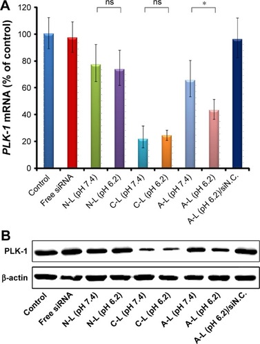
Figure 12 Schematic diagram of the proposed mechanism for A-L transportation across the cellular membrane and its delivery into cell cytoplasm.
Abbreviations: ACPP, activatable cell-penetrating peptide; A-L, ACPP-modified liposomes; pHe, extracellular pH.
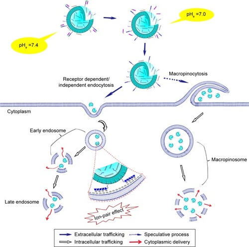
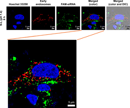
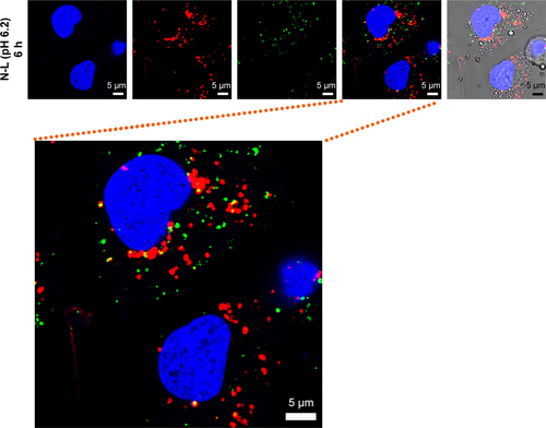
Figure S1 Intracellular trafficking and distribution of FAM-siRNA in MCF-7 cells.
Notes: The cells were treated with FAM-siRNA-encapsulated N-L at 37°C. The FAM-siRNA concentration was 350 nM. Cell nuclei and early endosomes were counterstained with Hoechst 33258 (blue) and CellLight Early Endosomes-RFP BacMam 2.0 (red), respectively. FAM-siRNA fluorescence (green) was recorded. Magnification 63×.
Abbreviations: DIC, differential interference contrast; FAM-siRNA, FAM-labeled small interfering RNA; N-L, nonmodified liposomes.