Figures & data
Figure 1 A) Rabbit model of interposition of external jugular vein into the common carotid artery using an end-to-side technique. B) Self-made drug-perfusion device.
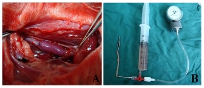
Figure 2 Histopathologic examination of vein grafts and normal vein with hematoxylin and eosin stain 28 days after operation (original magnification 200×). Bar = 100 μm. A) Group 1 (rapamycin-loaded PLGA nanoparticle vein graft perfusion), B) Group 2 (empty vehicle), C) Group 3 (vein graft with no treatment), and D) Group 4 (sham operation, normal vein).
Abbreviation: PGLA, poly lactic-co-glycolic acid.
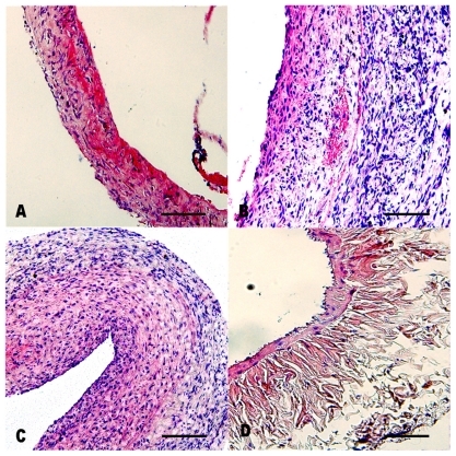
Figure 3 Pathologic examination of vein grafts and normal veins with Masson stain (original magnification 200×). A) Group 1 (rapamycin-loaded PLGA nanoparticle vein graft perfusion), B) Group 2 (empty vehicle), C) Group 3 (vein graft with no treatment), and D) Group 4 (sham operation, normal vein). In these figures, the collagen was dyed to red. Bar = 100 μm.
Abbreviation: PGLA, poly lactic-co-glycolic acid.
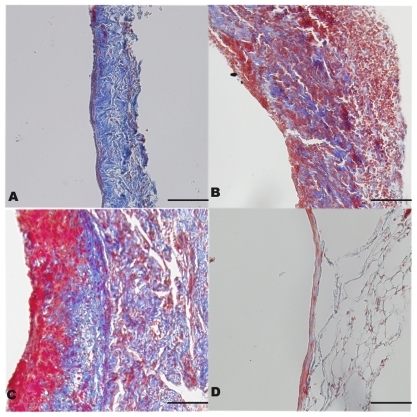
Figure 4 A) Intimal thickness of vein grafts and normal veins on postoperative day 28. *P < 0.05 compared with Group 4 and †P < 0.05 compared with Group 1. B) Ratio of intimal area to vessel area and collagen volume index in the four groups. *P < 0.05 compared with Group 3 and †P < 0.05 compared with Group 1. Data are expressed as mean ± standard deviation. C) Retention of rapamycin-loaded PLGA nanoparticles in the local vein graft at different doses. Differences between the two groups existed from seven to 28 days after treatment (P < 0.01).
Abbreviations: IA:VA, ratio of intimal area to vessel area; CVI, collagen volume index; PGLA, poly lactic-co-glycolic acid.
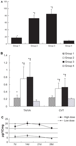
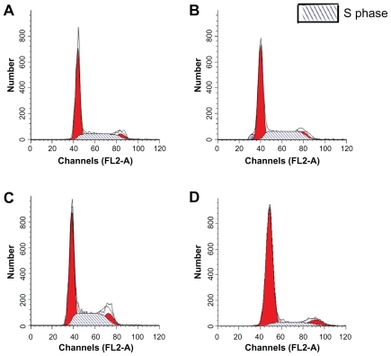
Figure 5 The flow cytometry assay for the proliferation cycles of the cells in the vein grafts and the normal vein. A) Group 1 (rapamycin-loaded PLGA nanoparticle vein graft perfusion), B) Group 2 (empty vehicle), C) Group 3 (vein graft with no treatment), and D) Group 4 (sham operation, normal vein). S phase, DNA synthesis phase.
Abbreviation: PGLA, poly lactic-co-glycolic acid.