Figures & data
Figure 1 A schematic illustration of preparation of PLA scaffold with microporous structure.
Abbreviations: EG, ethylene glycol; GC, THF, tetrahydrofuran; PLA, poly (d,l-lactic acid).
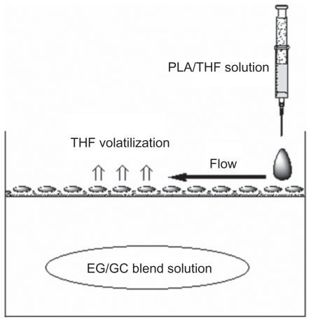
Figure 2 SEM of morphology of PLA film prepared via different methods. A) Volatilization naturally; film prepared via SNS phase separation at the concentration of B) 5%, C) 10%, and D) 15%. All samples were prepared at room temperature without air.
Abbreviations: PLA, poly(d,l-lactic acid); SEM, scanning electron microscope; SNS, solvent–nonsolvent.
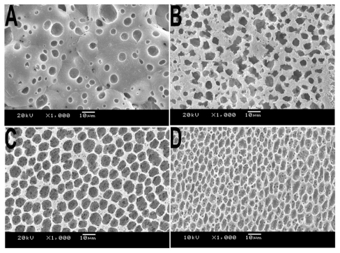
Figure 3 SEM micrographs of various types of PLA porous scaffold prepared at different temperatures. A) 4°C, B) 25°C, and C) 50°C. Inset: images of corresponding scaffold at higher magnification
Abbreviations: PLA, poly(d,l-lactic acid); SEM, scanning electron microscope.
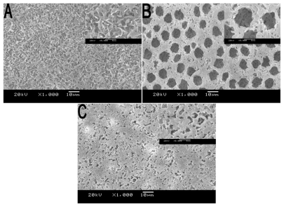
Figure 4 SEM images of PLA scaffolds prepared with various types of ventilation. A) Hermetic environment, B) open environment without air, and C) open environment with ventilated pumping equipment.
Abbreviations: PLA, poly(d,l lactic acid); SEM, scanning electron microscope.
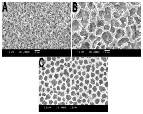
Figure 5 SEM images of PLA scaffolds prepared at 25°C with the concentration of 10% (w/v). Morphology observation of PLA film included the air face and cross-section: 1-a) air face of 10% PLA film, 1000×; 1-b) air surface of 10% PLA film, 3000×; 2-a) cross-section of 10% PLA film, 400×; 2-b) cross-section of 10% PLA film, 1000×.
Abbreviations: PLA, poly(d,l lactic acid); SEM, scanning electron microscope.
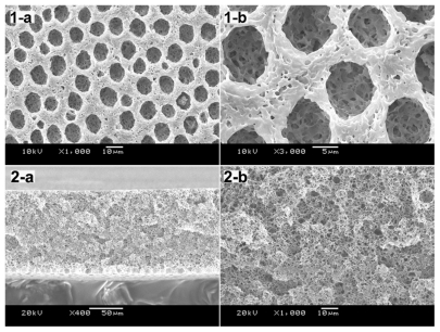
Table 1 Water contact angles and open porosity of the PLA scaffolds in different concentrations
Figure 6 SEM photographs showing morphology of NIH3T3 cells cultured on PLA scaffold. A) Cells cultured for 3 days, 200×; B) Cells cultured for 3 days, 2000×; C) Cells cultured for 12 h, 200×; D) Cells cultured for 12 h, 3500×.
Abbreviations: PLA, poly(d,l-lactic acid); SEM, scanning electron microscope.
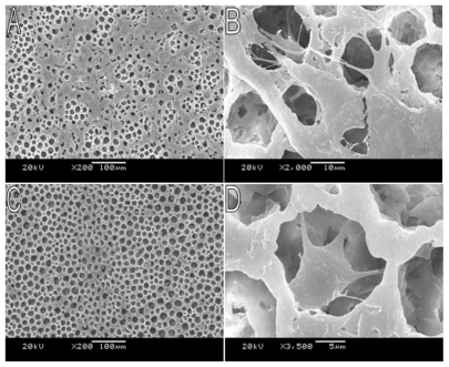
Figure 7 NIH3T3s proliferation on porous PLA scaffolds. The number of cells was normalized to initial density of seeded cells (1 × 10⁵ cells/well). Mean for n = 3 ± SD.
Abbreviations: PLA, poly(d,l-lactic acid); SD, standard deviation.
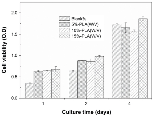
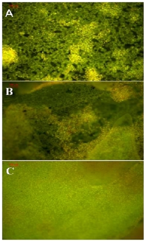
Figure 8 Fluorescence microscopy images of NIH3T3 cells cultured on A) 1, B) 2, and C) 4 days. The cells were stained by acridine orange stain.