Figures & data
Figure 1 Hemolytic activity of OVA-PD-Lipos in rabbit RBCs (n=3).
Abbreviations: OVA, ovalbumin; PD, Platycodin D; RBCs, red blood cells.
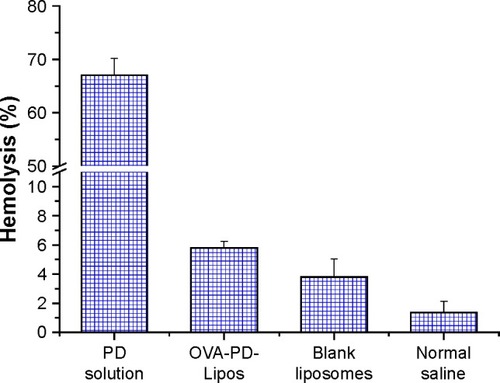
Figure 2 Toxicity of OVA-PD-Lipos to cultured mouse BMDCs (n=6).
Abbreviations: BMDCs, bone marrow dendritic cells; OVA, ovalbumin; PD, Platycodin D.
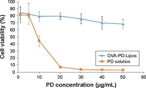
Figure 3 Uptake of FITC-OVA-PD-Lipos and FITC-OVA by cultured mouse BMDCs as determined by flow the units for the y-axes of this figure are cell count.
Abbreviations: BMDCs, bone marrow dendritic cells; FITC, fluorescein isothiocyanate; PD, Platycodin D; OVA, ovalbumin.
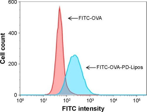
Figure 4 Transmission electron micrographs of (A) empty liposomes, (B) OVA-PD-Lipos, and (C) OVA-PD-Lipos-MNs.
Note: TEM was performed after OVA-PD-Lipos-MNs were dissolved in distilled water.
Abbreviations: OVA, ovalbumin; PD, Platycodin D; MNs, microneedle array.
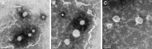
Figure 5 The integrity of OVA in OVA-PD-Lipos-MNs analyzed by SDS-PAGE.
Notes: Lane (A) protein molecular mass markers; Lane (B) OVA standard; Lane (C) OVA-PD-Lipos; Lane (D) OVA-PD-Lipos-MNs.
Abbreviations: OVA, ovalbumin; PD, Platycodin D; SDS-PAGE, sodium dodecyl sulfate polyacrylamide gel electrophoresis; MNs, microneedle array.
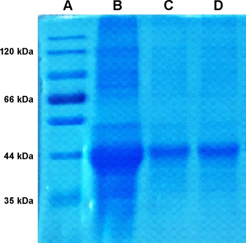
Figure 6 Evaluation of OVA-PD-Lipos-MNs inserted into mouse skin in vitro.
Notes: (A) Array of needle holes in mouse skin stained with trypan blue after insertion. (B and C) Vertical slices of mouse skin before and after insertion of OVA-PD-Lipos-MNs (stained using H&E).
Abbreviations: H&E, hematoxylin–eosin; OVA, ovalbumin; PD, Platycodin D; MNs, microneedle array.
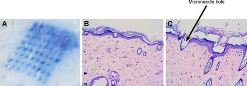
Figure 7 Images of nude mice after topical application of (A) FITC-OVA-PD-Lipos and (B) FITC-OVA-PD-Lipos-MNs. The colored area represents fluorescent intensity.
Abbreviations: FITC, fluorescein isothiocyanate; OVA, ovalbumin; PD, Platycodin D; MNs, microneedle array.
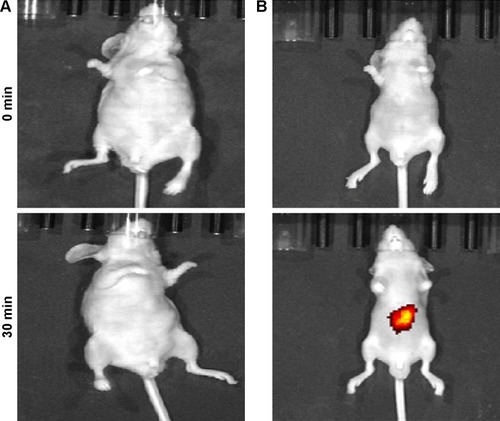
Figure 8 Dissolution of OVA-PD-Lipos-MNs after insertion into mouse skin in vivo.
Abbreviations: OVA, ovalbumin; PD, Platycodin D; MNs, microneedle array.
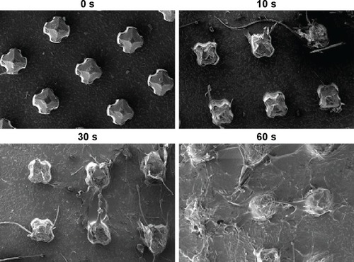
Figure 9 OVA-specific IgG, IgG1 and IgG2b levels in ICR mice immunized with OVA-PD-Lipos-MNs (n=6).
Notes: OVA, OVA + PD, and OVA-PD-Lipos represent OVA solution, OVA and PD solution, OVA and PD loaded liposomes, respectively; OVA-MNs, OVA-PD-MNs, and OVA-PD-Lipos-MNs represent free OVA, free OVA + PD, and OVA-PD-Lipos, loaded into dissolving microneedle array, separately; no statistical difference was found among groups in the same box; *P<0.05.
Abbreviations: OVA, ovalbumin; PD, Platycodin D; MNs, microneedle array.
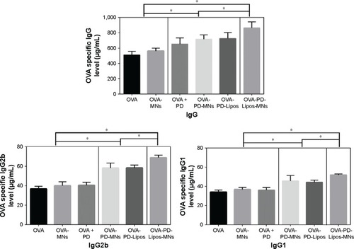
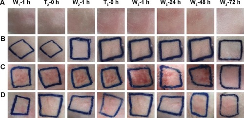
Figure 10 Response of intact rabbit skin after multiple-dose delivery of OVA-PD-Lipos-MNs.
Notes: Rows A–D represent blank control, blank dissolving microneedles, free OVA and PD in dissolving microneedles, and OVA-PD-Lipos-MNs, respectively. Column W1-1 h and W2-1 h represent 1 h after the washing out of the first and second dose; Column W3-1 h, W3-24 h, W3-48 h, and W3-72 h represent 1, 24, 48, and 72 h after the washing out of the third dose, respectively. Column T2-0 h and T3-0 h represent immediately before the application of the second and third dose.
Abbreviations: OVA, ovalbumin; PD, Platycodin D; MNs, microneedle array.