Figures & data
Figure 1 Gold nanoparticle (AuNP) synthesis and characterization.
Notes: (A) Transmission electron micrography of spherical monodispersed citrate-stabilized AuNPs (12±3 nm); (B, C) Ultraviolet-visible absorbance spectra of AuNP stability after modification with PVP and mPEG in physiological salt solution (PSS) and culture media.
Abbreviations: PVP, polyvinylpyrrolidone; mPEG, mercapto polyethylene glycol.
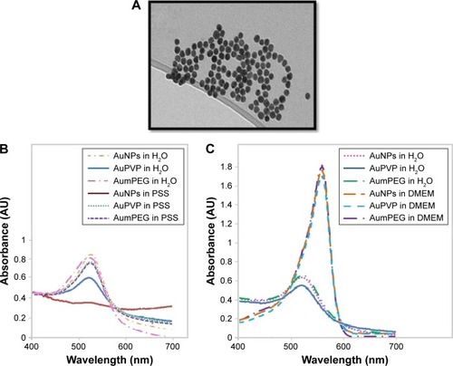
Figure 2 FTIR spectra for stabilizers and unmodified and modified AuNPs.
Note: (A) PVP, (B) AuPVP, (C) mPEG, (D) AumPEG, and (E) AuNPs, illustrating characteristic absorption peaks.
Abbreviations: FTIR, Fourier-transform infrared spectroscopy; AuNPs, gold nanoparticles; PVP, polyvinylpyrrolidone; mPEG, mercapto polyethylene glycol.
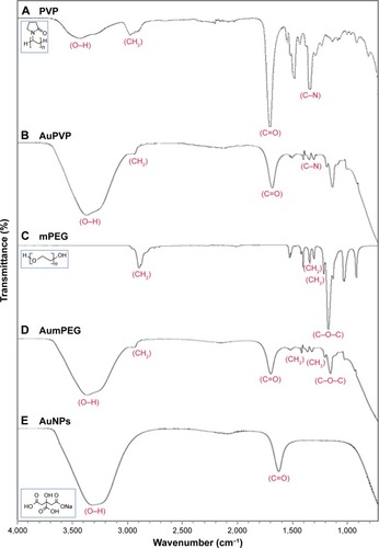
Figure 3 Gold nanoparticle (AuNP) uptake by BAECs in culture.
Notes: Transmission electron micrographs showing the time course of AuNP uptake (citrate-stabilized and mPEG- and PVP-modified) by BAECs. AuNPs are identified as spherical structures inside endosomes within the endothelial cells (arrows).
Abbreviations: BAECs, bovine aortic endothelial cells; mPEG, mercapto polyethylene glycol; PVP, polyvinylpyrrolidone.
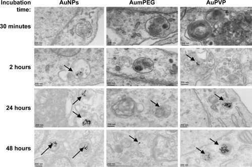
Figure 4 Inhibitory effects of stabilizers and unmodified and modified AuNPs on BAEC proliferation.
Notes: **P<0.01; ***P<0.001. Results presented as mean ± SD. Three independent experiments.
Abbreviations: AuNPs, gold nanoparticles; BAEC, bovine aortic endothelial cell; mPEG, mercapto polyethylene glycol; PVP, polyvinylpyrrolidone.
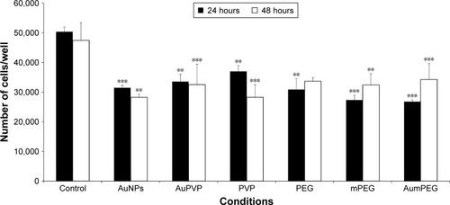
Figure 5 The influence of stabilizers and unmodified and modified AuNPs on BAEC viability using flow cytometry.
Notes: *P<0.05; **P<0.01. Percentage cell viability of BAECs in the untreated cells and cells treated with stabilizer (PVP), non-modified and modified AuNPs (AuPVP, AumPEG) after 30-minute and 2–24- and 48-hour exposure based on three independent experiments. Results presented as mean ± SD.
Abbreviations: AuNPs, gold nanoparticles; BAEC, bovine aortic endothelial cell; PVP, polyvinylpyrrolidone; mPEG, mercapto polyethylene glycol.
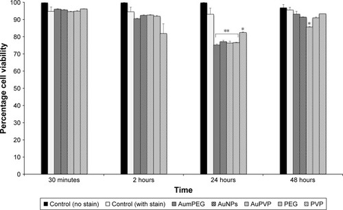
Figure 6 Induction of apoptosis and necrosis by stabilizers and unmodified and modified AuNPs in BAECs.
Notes: *P<0.05; **P<0.01; ***P<0.001. Apoptosis (A) and necrosis (B) of BAECs in untreated cells and cells treated with stabilizer (PVP) and unmodified and modified AuNPs (AuPVP, AumPEG) after 2- and 24-hour exposure based on three independent experiments. Results presented as mean ± SD.
Abbreviations: AuNPs, gold nanoparticles; BAECs, bovine aortic endothelial cells; PVP, polyvinylpyrrolidone; mPEG, mercapto polyethylene glycol.
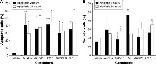
Figure 7 Modulatory effects of stabilizer and unmodified and modified AuNPs on ACh-induced ERK1/2 phosphorylation in BAECs.
Notes: (A) Representative Western blot analysis (A1) showing the stimulatory effect of 1/10/100 µM ACh on ERK1/2 phosphorylation (p-ERK1/2) in BAECs after 10 minutes’ exposure compared to untreated cells (control); relative expression of p-ERK1/2 calculated as a ratio to ERK1/2 expression, the loading control (A2). (B) Representative Western blot analysis (B1) showing the modulatory effects of 10 µM ACh-induced ERK1/2 phosphorylation in the absence or in the presence of stabilizer (PVP) and non-modified and unmodified AuNPs (AuPVP, AumPEG) in BAECs after 10 minutes’ incubation compared to untreated cells (control). The bar graph shows the relative expression of p-ERK1/2 calculated as a ratio to ERK1/2 expression, the loading control (B2).
Abbreviations: AuNPs, gold nanoparticles; ACh, acetylcholine; BAECs, bovine aortic endothelial cells; PVP, polyvinylpyrrolidone; mPEG, mercapto polyethylene glycol; t-ERK, total ERK.
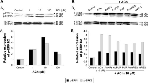
Figure 8 Gold nanoparticle (AuNP) uptake by aortic vessels.
Notes: (A) Determination of AuNP uptake using inductively coupled plasma analysis. Concentration of unmodified and polymer-modified AuNPs within aortic vessels after 30 minutes’ exposure. Error bars are SEM. (B) Representative transmission electron micrography illustrating the uptake of PVP-modified AuNPs into endosomal structures (a; b at higher magnification), and also on the surface of endothelial cells lining the aortic vessel (c), within 30 minutes’ exposure. Very few mPEG-modified AuNPs are seen as spherical dense structures within the endothelial cells (d, arrow) when exposed to the higher concentration.
Abbreviations: PVP, polyvinylpyrrolidone; mPEG, mercapto polyethylene glycol.
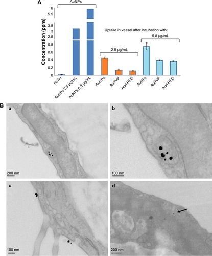
Figure 9 Influence of stabilizers and unmodified and modified AuNPs on dilator responses of aortic vessels.
Notes: *P<0.05; **P<0.01; ***P<0.001. Endothelial-dependent (ACh) and independent (SNP) dilator responses of preconstricted aortic vessels after 30 minutes’ exposure ex vivo. Error bars are SEM.
Abbreviations: AuNPs, gold nanoparticles; ACh, acetylcholine; SNP, sodium nitroprusside; PVP, polyvinylpyrrolidone; mPEG, mercapto polyethylene glycol.
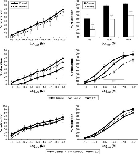
Figure S1 Surface-enhanced Raman spectroscopy analysis for unmodified and modified AuNPs.
Note: Unmodified AuNPs (A), mPEG-modified AuNPs (B), and PVP-modified AuNPs (C).
Abbreviations: AuNPs, gold nanoparticles; mPEG, mercapto polyethylene glycol; PVP, polyvinylpyrrolidone.

Figure S2 Flow-cytometry analysis of cell viability, apoptosis, and necrosis after AuNP exposure.
Notes: (A) Cell viability of BAECs in untreated cells and in cells treated with AumPEG after 24 and 48 hours. Numbers within dot plots represent the percentage of live cells (lower left, PI−). (B) Induction of apoptosis by AumPEG after 2 hours’ exposure, showing induction of necrosis by stabilizers after 2 and 24 hours’ exposure in comparison with untreated cells.
Abbreviations: AuNP, gold nanoparticle; BAECs, bovine aortic endothelial cells; mPEG, mercapto polyethylene glycol; PI, propidium iodide; PE, phycoerythrin; PVP, polyvinylpyrrolidone.
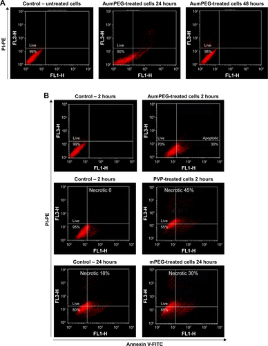
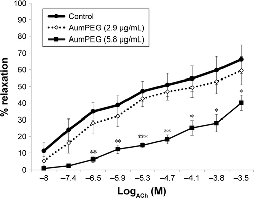
Figure S3 Influence of mPEG-modified AuNPs on endothelial-dependent dilator responses of aortic vessels.
Notes: *P<0.05; **P<0.01; ***P<0.001. Endothelial-dependent (ACh) dilator responses of preconstricted aortic vessels after 30 minutes’ exposure of high-concentration mPEG-modified AuNPs (5.8 µg/mL) ex vivo. Error bars are SEM.
Abbreviations: AuNPs, gold nanoparticles; mPEG, mercapto polyethylene glycol; PVP, polyvinylpyrrolidone.