Figures & data
Figure 1 Strategy of exosome pseudotyping.
Notes: (A) Membrane topology of the fVSVG. The matured fVSVG is a single transmembrane protein without the SP. The large N-terminal ectodomain (black line) with a short stem region (green line) is situated at the outer surface of the plasma membrane or the luminal side of the endosome. Those sequences are followed by a transmembrane helix and a cytoplasmic tail. (B) Design of VSVG fusion constructs. From top to bottom, the fVSVG fused with either RFP (fVSVG-RFP) or GFP (fVSVG-GFP) at the C-terminal, the ectodomain was replaced by RFP (RFP-mVSVG), the fVSVG fused with Gaussia luciferase (Gluc) at the C-terminal (fVSVG-Gluc), and the ectodomain was replaced by Gluc (Gluc-mVSVG). (C) A proposed model illustrating how VSVG participates in exosomes in a mammalian cell. Ectopic expression of VSVG occurs at the rough endoplasmic reticulum (ER) via its SP guiding, and subsequently SP-cleaved VSVG is funneled to the plasma membrane and becomes concentrated in tetraspanin (CD63)-enriched microdomains, where the first inward budding begins to form endosomes. The second inward budding from the endosome forms exosomes that are stored in a MVB prior to release into extracellular space. (D) Schematic illustration of the pseudotyped and protein-loaded exosome. Membrane topology of VSVG (black), loaded protein cargo GFP (green) or RFP (red), and exosome markers (CD63/CD81, blue) are indicated.
Abbreviations: VSVG, vesicular stomatitis virus glycoprotein; fVSVG, full-length VSVG; mVSVG, minimal VSVG; MVB, multiple-vesicle body; SP, signal peptide.
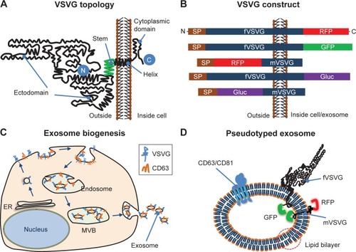
Figure 2 Fluorescent imaging of VSVG fusion proteins in HEK293 cells.
Notes: Cultured cells were transfected with either fVSVG-GFP/fVSVG-RFP alone or in combination with Rab5A-GFP/CD63-GFP for indicated periods of time. Cell images of fluorescence signal and phase contract of the same field were taken to show the expression and an earlier plasma membrane distribution of fVSVG-GFP on day 2 (A, green; B, overlay), and late-punctate intracellular localization on day 3 (C, green; D, overlay). Similarly, the expression and subcellular localization of fVSVG are shown in red (E–H). Interestingly, following cotransfection of cells with fVSVG-RFP and Rab5A-GFP (an endosome marker) for 3 days, images show the expression and cellular distribution of fVSVG (I, red), Rab5A (J, green), or colocalization of both (K, yellow). Alternatively, cotransfection with both fVSVG-RFP and CD63-GFP (an exosome marker) resulted in similar patterns of expression and cellular distribution for fVSVG (L, red), CD63 (M, green), and colocalization of both (N, yellow). Arrows indicate endosome/exosome/MVB structures. Scale bar 20 µm.
Abbreviations: VSVG, vesicular stomatitis virus glycoprotein; fVSVG, full-length VSVG; MVB, multiple-vesicle body; SP, signal peptide.
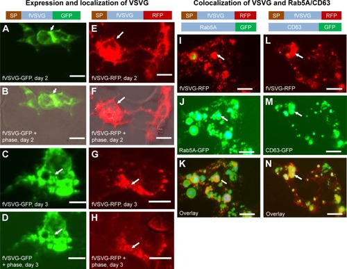
Figure 3 Validation of minimal VSVG (mVSVG) scaffold for exosome targeting.
Notes: HEK293 cells were cotransfected with RFP-mVSVG and fVSVG-GFP for 3 days. Cell images were taken by fluorescence microscopy to show the intracellular expression of mVSVG (A, red), fVSVG (B, green), and colocalization of both (C, yellow). Alternatively, cells were cotransfected with RFP-mVSVG and endosome marker Rab5A-GFP for 3 days. Expression and subcellular distribution were shown for mVSVG (D, G; red), Rab5A (E, green) and colocalization of both in overlay (F, yellow). Similar results were obtained to show the expression and subcellular distribution of exosome marker CD63 (H) and its colocalization with mVSVG (I, yellow). Arrows indicate endosome/exosome/MVB structures. Scale bar 20 µm.
Abbreviations: VSVG, vesicular stomatitis virus glycoprotein; fVSVG, full-length VSVG; mVSVG, minimal VSVG; MVB, multiple-vesicle body; SP, signal peptide.
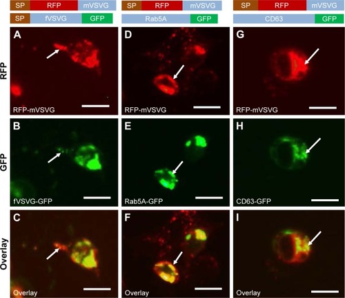
Figure 4 Time course of VSVG fusion incorporation into exosomes.
Notes: HEK293 cells were transfected with fVSVG-RFP or XPack-GFP alone (an exosome tracer) for indicated periods of time. The incorporation of fVSVG into exosomes was monitored and compared with that of XPack. Live cell images were taken at days 1, 2, 4, and 6; representative images are shown to illustrate the expression of fVSVG (A, top panel) and XPack (A, bottom panel) at early membrane appearance (days 1 and 2), and final incorporation in exosomes (days 4 and 6). (B) In a separate set of experiments, cells were cotransfected with fVSVG-RFP and XPack-GFP for 3 days. The expression of fVSVG (red), XPack (green), and colocalization of both (yellow in overlay) are shown. Arrows indicate membrane locations and endosome/exosome/MVB structures. Scale bar 20 µm.
Abbreviations: VSVG, vesicular stomatitis virus glycoprotein; mVSVG, minimal VSVG; fVSVG, full-length VSVG; MVB, multiple-vesicle body.
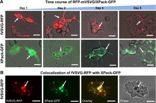
Figure 5 Secretion and characterization of pseudotyped exosomes.
Notes: (A) Configuration of Gaussia luciferase reporters used for monitoring exosome secretion. (B) Gaussia luciferase activity was assayed using conditioned media from HEK293 cell culture on day 2 posttransfection, with mock transfection as negative controls. Data presented as relative light units from three experiments (mean ± standard deviation, n=3). (C) Immunological pull-down of pseudotyped exosomes using CD81-specific antibody (Ab)-coated beads. Both images of green fluorescence and phase contrast are displayed to show GFP-positive anti-CD81 antibody-precipitated exosomes. Non-Ab-coated beads were included as controls. Arrows indicate exosome-positive beads. (D–F) Nanoparticle-tracking analysis profiles off VSVG, mVSVG, or CD63 pseudotyped exosomes isolated from HEK293 cells at 3 days posttransfection, showing average sizes of respective exosomes. (G) There was no difference in average exosome size between pseudotyped fVSVG-RFP/RFP-mVSVG and modified CD63 (an endogenous exosome marker).
Abbreviations: VSVG, vesicular stomatitis virus glycoprotein; mVSVG, minimal VSVG; fVSVG, full-length VSVG; SP, signal peptide.
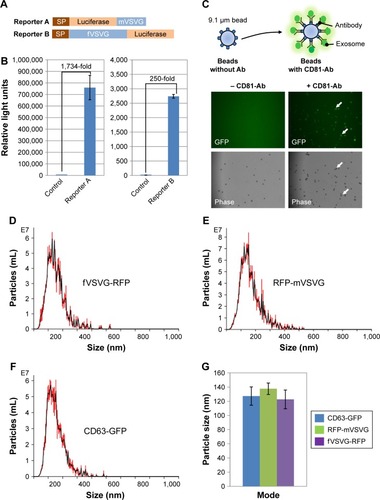
Figure 6 Exosome uptake in mammalian cells.
Notes: Uptake of pseudotyped exosomes in various cell lines (HEPG2, U97, HEK293, L929, iPSC11, and iPSC15) was compared between fVSVG and mVSVG. Cultured cells were treated with either fVSVG-RFP or RFP-mVSVG in a 96-well plate. Enhanced uptake of fVSVG was evident (A–C and G–K) compared to mVSVG (D–F and H–L). Following exosome incubation for 48 hours, images were taken with fluorescent microscopy at 20× magnification for HEPG2, U97, HEK293, and L929 cells (scale bar 20 µm). For iPSC11 and iPSC15 cells, images were taken after exosome incubation for 24 hours at 10× magnification (scale bar 10 µm). Arrows point to internalized RFP-labeled exosomes.
Abbreviations: VSVG, vesicular stomatitis virus glycoprotein; fVSVG, full-length VSVG; mVSVG, minimal VSVG.
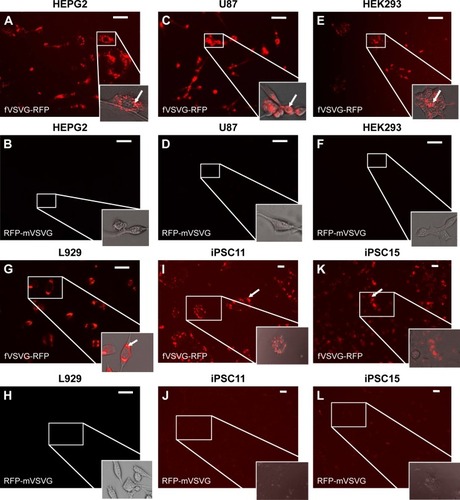
Figure 7 Quantification of exosome uptake in HEK293 cells by flow cytometry.
Notes: Recipient cells were loaded with either fVSVG-RFP or RFP-mVSVG pseudotyped exosomes for 48 hours, then washed and subjected to fluorescence-activated cell-sorting analysis. Right shifts in fluorescence signals for both fVSVG (C) and mVSVG (B) exosomes are shown in comparison with the negative control (A) in panel (D), indicating an enhancement (~9.7-fold) in exosome uptake by pseudotyping in HEK293 cells.
Abbreviations: VSVG, vesicular stomatitis virus glycoprotein; fVSVG, full-length VSVG; mVSVG, minimal VSVG.
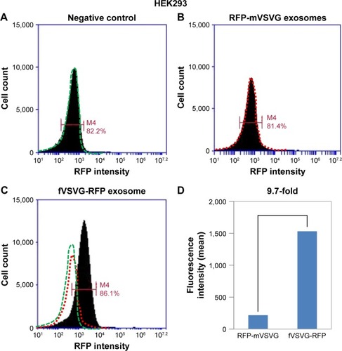
Figure S1 Colocalization of mVSVG with exosome marker CD81 in HEK293 cells.
Notes: Cultured cells were cotransfected with RFP-mVSVG and exosome marker CD81-GFP for 3 days. Cell images of the same field were taken to show the expression and subcellular localization of mVSVG (A, red), CD81 (B, green), and colocalization of both (C, yellow). Arrows indicate endosome/exosome/MVB structures. Scale bar 20 µm.
Abbreviations: VSVG, vesicular stomatitis virus glycoprotein; mVSVG, minimal VSVG; MVB, multiple-vesicle body; SP, signal peptide.
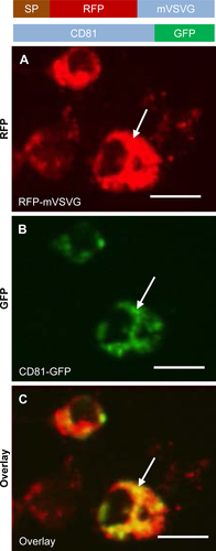
Figure S2 Intracellular localization of VSVGs in multiple mammalian cell lines.
Notes: Cultured cells were transfected with either fVSVG-RFP or RFP-mVSVG for 3 days in U87, HEPG2, and L292 lines. Images of fluorescence signal and phase contract of the same field were taken to show the expression and subcellular localization of fVSVG (A) or mVSVG (B). Arrows and overlay indicate endosome/exosome/MVB structures inside cells. Scale bar 20 µm.
Abbreviations: VSVG, vesicular stomatitis virus glycoprotein; fVSVG, full-length VSVG; mVSVG, minimal VSVG; MVB, multiple-vesicle body; SP, signal peptide.
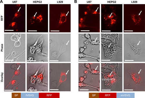
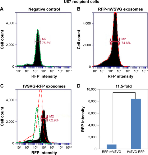
Figure S3 Quantification of exosome uptake in U87 cells by flow cytometry.
Notes: Cells at ~30% confluence were loaded with pseudotyped exosomes in a six-well plate. After 48 hours of incubation, cells were washed and subjected to fluorescence-activated cell-sorting analysis. Right shifts in fluorescence signals for both fVSVG-RFP (C) and RFP-mVSVG (B) exosomes are shown in comparison with the negative control (A), indicating an enhancement (~11.5-fold) in exosome uptake by pseudotyping in U87 recipient cells (D).
Abbreviations: VSVG, vesicular stomatitis virus glycoprotein; fVSVG, full-length VSVG; mVSVG, minimal VSVG.