Figures & data
Figure 1 FT-IR spectrum of O-CNT and O-CNT-PEG.
Abbreviations: au, absorbance units; FT-IR, Fourier Transform infrared; O-CNT, oxidized carbon nanotube; PEG, polyethylene glycol.
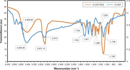
Figure 2 TGA thermo grams of O-CNT and O-CNT-PEG.
Abbreviations: au, absorbance units; O-CNT, oxidized carbon nanotube; PEG, polyethylene glycol; TGA, thermogravimetric analysis.
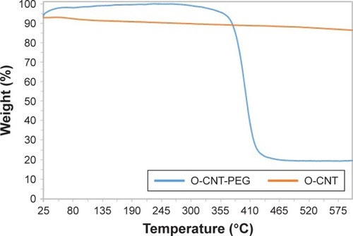
Figure 3 FESEM images of functionalized CNTs.
Notes: (A) O-CNT and (B) O-CNT-PEG.
Abbreviations: FESEM, field emission scanning electron microscopy; O-CNT, oxidized carbon nanotube; PEG, polyethylene glycol; CNT, carbon nanotubes.
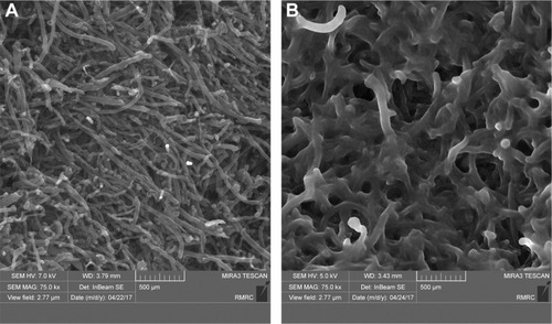
Figure 4 UV–Vis absorption spectrum of O-CNT-PEG.
Abbreviations: au, absorbance units; O-CNT, oxidized carbon nanotube; PEG, polyethylene glycol; UV–Vis, ultraviolet–visible.
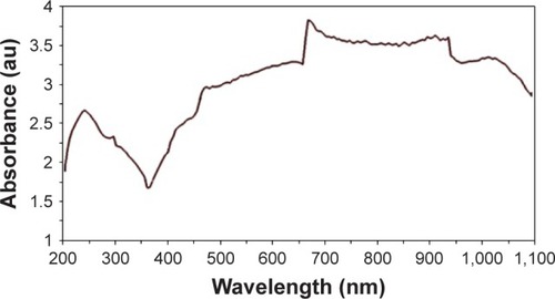
Figure 5 MTT assay of MWNT. O-CNT, and O-CNT-PEG against HepG2 (A) and HeLa (B) cell lines.
Abbreviations: Conc, concentration; MWNT, multiwalled carbon nanotube; O-CNT, oxidized carbon nanotube; PEG, polyethylene glycol.
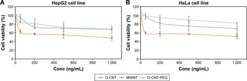
Figure 6 The size of tumors before and 3 days after the treatment with PTT in different groups.
Abbreviations: O-CNT, oxidized carbon nanotube; PEG, polyethylene glycol; PTT, photothermal therapy.
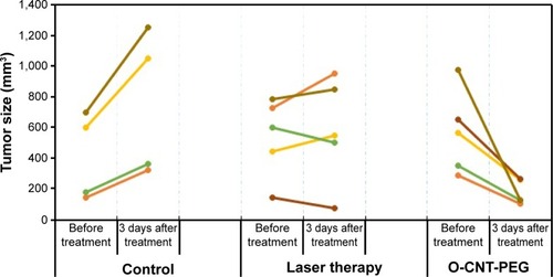
Figure 7 The stages of tumor treatment in the O-CNT-PEG group with PTT technique.
Notes: (A, C) Photograph and ultrasonography image of a cancerous mouse before the treatment, respectively. (B) Photograph of the mouse 3 days after the treatment (sonography was not feasible 3 days after the treatment).
Abbreviations: O-CNT, oxidized carbon nanotube; PEG, polyethylene glycol; PTT, photothermal therapy.
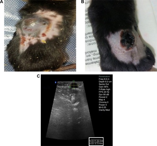
Table 1 Results of histopathologic evaluation
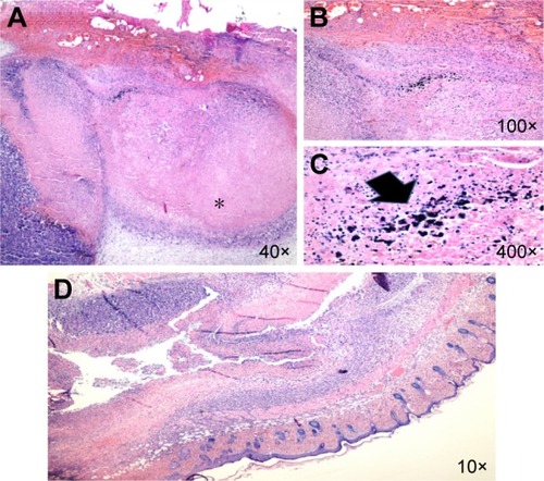
Figure 8 Malignant melanoma.
Notes: (A) (*) Histopathologic section of the laser therapy case which shows the zone of tumor necrosis (H&E). (B, C) There is the deposition of NPs (arrow) in O-CNT-PEG group within the necrotic areas (H&E). (D) Histopathologic section of the tumor in the case allocated to the O-CNT-PEG group which shows high percent of tumor necrosis (10×; (H&E)).
Abbreviations: H&E, hematoxylin and eosin; NPs, nanoparticles; O-CNT, oxidized carbon nanotube; PEG, polyethylene glycol.