Figures & data
Table 1 Characteristics of the PGA and PGG polymers and the PGG-PTX
Figure 1 Synthesis of PGG-PTX nanoconjugate.
Abbreviations: HOBt, hydroxybenzotriazole; TFA, atrifluoroacetic acid; DMAP, 4-dimethylaminopyridine; NaHCO3, sodium bicarbonate; PGA, poly(L-glutamic acid); PGG, poly(L-γ-glutamyl-glutamine); PGG-PTX, poly(L-γ-glutamyl-glutamine)-paclitaxel conjugate.
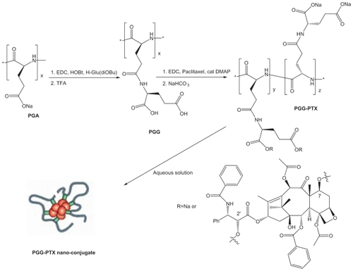
Figure 2 SEC-HLPC chromatograms of PGA, PGG, and PGG-PTX. The chromatograms were recorded at 228 nm. A) PGA; B) PGG; C) PGG-PTX.
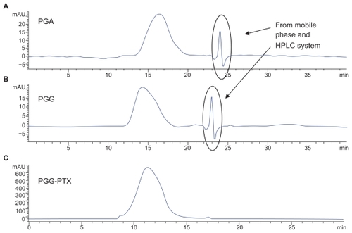
Figure 3 Critical micellar concentration of PGG-PTX nanoconjugate. The DLS could not detect the particle size of PGG-PTX solution below 25 μg/mL. The critical micellular concentration was assumed to be about 25 μg/mL in saline at 25°C. The results are expressed as means ± SD (n = 3).
Abbreviation: DLS, dynamic light scattering.
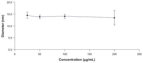
Figure 4 Photographs of a solution of A) PGG-PTX (35% PTX loading) and B) PGA-PTX (32% PTX loading) in saline (0.9% NaCl). The polymer-PTX conjugates were dissolved in 0.9% NaCl at 50 mg/mL after sonication for 1 minute and allowed to stand for 20 minutes.
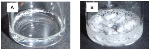
Figure 5 Inhibition of the growth of human lung cancer H460 cells as a function of concentration of PTX (O), PGA-PTX (■) and (▴) PGG-PTX.
Note: Vertical bars, SEM.
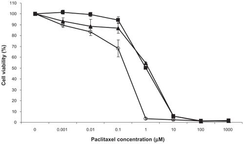
Figure S1 1H-NMR spectra of PGA-PGG, and PGG-PTX.
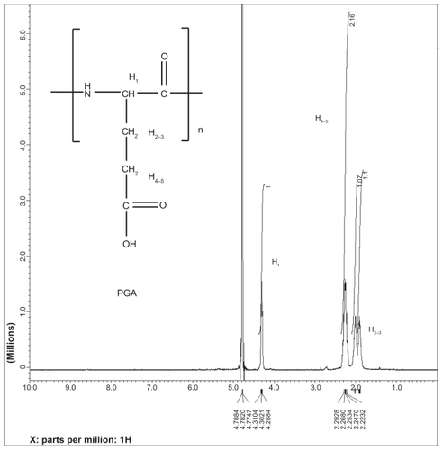
Figure S2 1H-NMR spectra of PGA, PGG, and PGG-PTX.
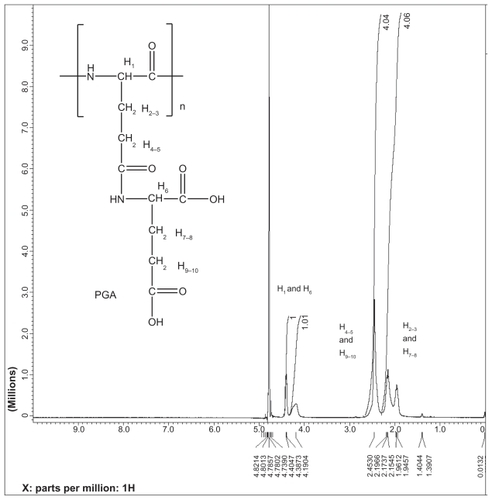
Figure S3 PGG and PGG-PTX chromatgrams using light scattering and refractive index detectors.
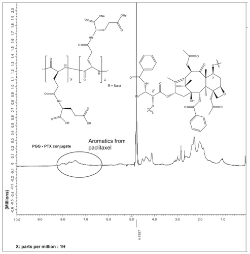
Figure S4 Red line came from light scattering detector. Blue line came from refractive index.
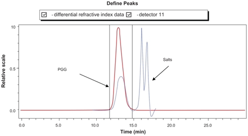
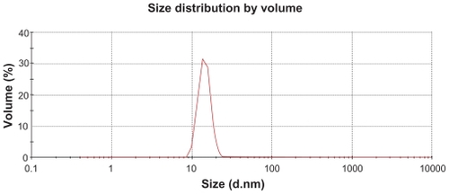
Figure S5 Size distribution of PGG-PTX in saline with various concentrations
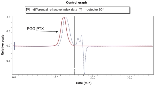
Figure S6 PGG-PTX (2,000 μg/mL) in saline. Diameter = 13.7 nm; PDI = 0.404.
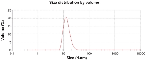
Figure S7 PGG-PTX (50 μg/mL) in saline. Diameter = 14.7 nm; PDI = 0.599.