Figures & data
Figure 1 Schematic illustration of the synthesized silver/montmorillonite/chitosan bionanocomposites from silver nitrate/montmorillonite/chitosan (A0) by ultraviolet irradiation method.
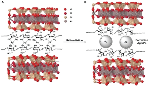
Figure 2 Photograph of montmorillonite, silver nitrate/montmorillonite/chitosan (A0), silver/montmorillonite/chitosan bionanocomposites (A2, A4, and A5) for 3, 48, and 96 hours of ultraviolet irradiation times.
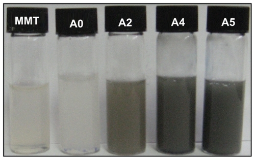
Figure 3 Ultraviolet-visible adsorption spectra of silver nitrate/montmorillonite/chitosan (A0) and silver/montmorillonite/chitosan bionanocomposites (A1–A5) at different ultraviolet irradiation times.

Figure 4 Powder X-ray diffraction, patterns of montmorillonite, montmorillonite/chitosan, and silver/montmorillonite/chitosan bionanocomposites for (A2) three hours, (A4), 48 hours, and (A5) 96 hours of ultraviolet irradiation times.
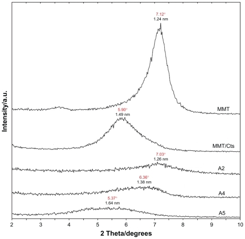
Figure 5 Powder X-ray diffraction patterns of montmorillonite, silver/montmorillonite/chitosan bionanocomposites for different ultraviolet irradiation times (A1) 1 hour, (A2) 3 hours, (A3) 18 hours, (A4) 48 hours, and (A5) 96 hours.
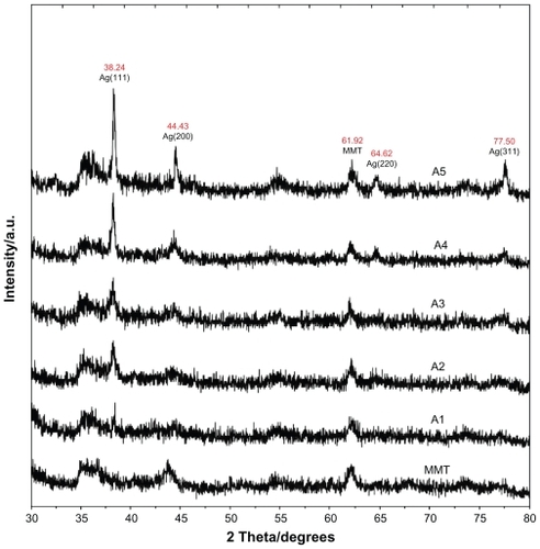
Figure 6 Transmission electron microscopy images and their corresponding particle size distributions of silver/montmorillonite/chitosan bionanocomposites for A2 (A, B), A4 (C, D), and A5 (E, F).
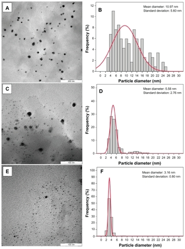
Figure 7 Scanning electron microscopy micrographs and energy dispersive x-ray fluorescence spectra, respectively, for the montmorillonite (A, B), montmorillonite/chitosan (C, D) and silver/montmorillonite/chitosan bionanocomposites [A5 (E, F)].
![Figure 7 Scanning electron microscopy micrographs and energy dispersive x-ray fluorescence spectra, respectively, for the montmorillonite (A, B), montmorillonite/chitosan (C, D) and silver/montmorillonite/chitosan bionanocomposites [A5 (E, F)].](/cms/asset/04409d4a-78fa-40b7-ad87-3b6ef3e9af31/dijn_a_12184750_f0007_c.jpg)
Figure 8 Fourier transform infrared spectra for A) chitosan, B) montmorillonite/chitosan, and C) montmorillonite.
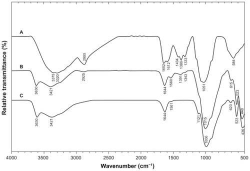
Figure 9 Fourier transform infrared spectra of A) silver/montmorillonite/chitosan bionanocomposites for (A5) and B) montmorillonite/chitosan.
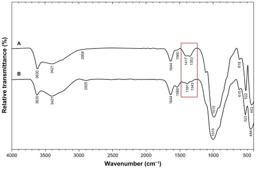
Figure 10 Comparison of the inhibition zone test between montmorillonite, silver nitrate/montmorillonite/chitosan (A0), and silver/montmorillonite/chitosan bionanocomposites (A2, A4, and A5) against different bacteria.
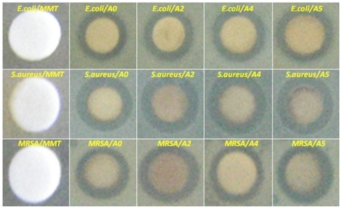
Table 1 Average inhibition zone and standard deviation for Ag/montmorillonite/chitosan bionanocomposites (A2, A4, and A5) and AgNO3/montmorillonite/chitosan (A0)