Figures & data
Figure 1 Synthesis scheme of PEG-b-PLMA and Gal-PEG-b-PLMA copolymers.
Abbreviations: PEG-b-PLMA, methoxy poly(ethylene glycol)/poly(l-lactide-co-β-malic acid) block copolymer; Gal-PEG-b-PLMA, galactosylated methoxy poly(ethylene glycol)/poly(l-lactide-co-β-malic acid) block copolymer.
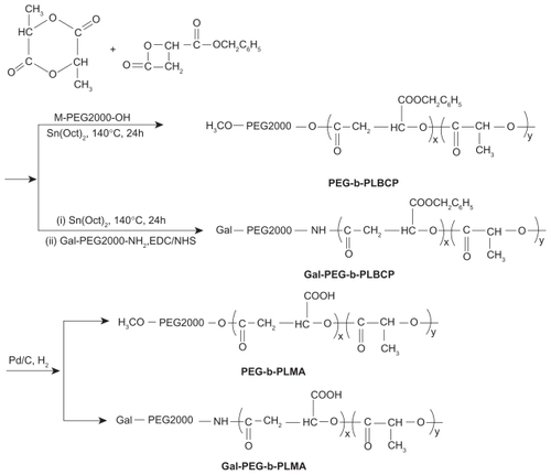
Table 1 Physical properties of PLMA and PEG-b-PLMA copolymers
Figure 2 Size distributions of (A) empty, (B) doxorubicin-loaded Gal-PEG-b-PLMA micelles, and (C) their mean sizes measured by dynamic light scattering method.
Abbreviation: Gal-PEG-b-PLMA, galactosylated methoxy poly(ethylene glycol)/poly(l-lactide-co-β-malic acid) block copolymer.
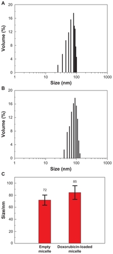
Figure 3 Transmission electron microscopic images of (A) empty and (B) doxorubicin loaded Gal-PEG-b-PLMA micelles.
Abbreviation: Gal-PEG-b-PLMA, galactosylated methoxy poly (ethylene glycol)/poly (l-lactide-co-β-malic acid) block copolymer.
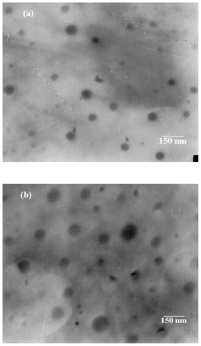
Figure 4 In vitro release behavior of doxorubicin from PEG-b-PLMA micelles at pH 4.5, 5.5, and 7.4. Each point and error bar represents mean ± standard deviation (n ± 3).
Abbreviation: PEG-b-PLMA, methoxy poly(ethylene glycol)/poly(l-lactide-co-β-malic acid).
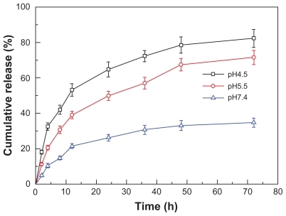
Figure 5 In vitro cytotoxicity of free doxorubicin, doxorubicin-loaded PEG-b-PLMA micelles and doxorubicin-loaded Gal-PEG-b-PLMA micelles in HepG2 cells. Data are shown as mean ± standard deviation (n = 3).
Abbreviations: PEG-b-PLMA, methoxy poly(ethylene glycol)/poly(l-lactide-co-β-malic acid); Gal-PEG-b-PLMA, galactosylated methoxy poly(ethylene glycol)/poly (l-lactide-co-β-malic acid).
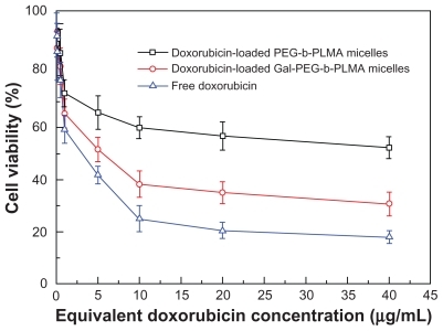
Figure 6 The cell cycle profiles of HepG2 cells before and after doxorubicin treatment for 12 hours: A) control group, B) free doxorubicin group, C) doxorubicin-loaded PEG-b-PLMA micelle group, and D) doxorubicin-loaded Gal-PEG-b-PLMA micelle group. The data represent mean ± standard deviation where n = 3.
Abbreviations: PEG-b-PLMA, methoxy poly(ethylene glycol)/poly(l-lactide-co-β-malic acid); Gal-PEG-b-PLMA, galactosylated methoxy poly(ethylene glycol)/poly (l-lactide-co-β-malic acid).
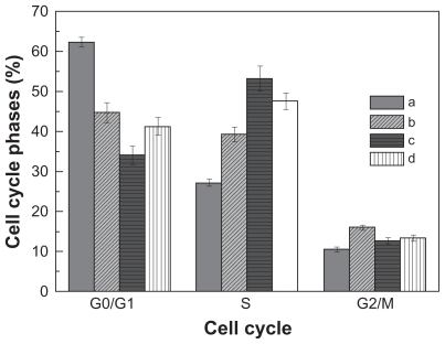
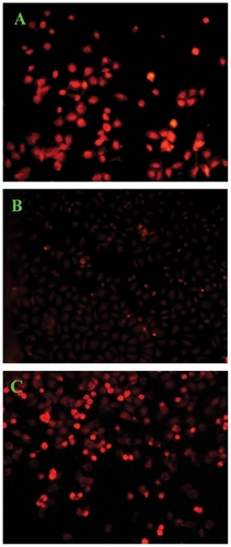
Figure 7 Confocal laser scanning microscopic images of HepG2 cells (5 × 10⁴ cells/mL) after 12 hours of in vitro exposure to (A) free doxorubicin, (B) doxorubicin-loaded PEG-b-PLMA micelles, and (C) doxorubicin-loaded Gal-PEG-b-PLMA micelles.
Abbreviations: PEG-b-PLMA, methoxy poly(ethylene glycol)/poly (l-lactide-co-β-malic acid); Gal-PEG-b-PLMA, galactosylated methoxy poly(ethylene glycol)/poly(llactide-co-β-malic acid).