Figures & data
Table 1 Type, chemical composition, and size of particles identified in lymph node samplesTable Footnote*
Figure 1 The microphotograph shows two submicronic particles (the white spots) embedded in the left lymph-node biopsy sample. The EDS spectrum shows the chemical composition: carbon, oxygen, chromium, phosphorus, sulfur, and chlorine.
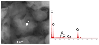
Figure 2 The microphotograph shows the analyses carried out on the right lymph-node with the EDS spectrum on the chemical composition of the debris. Nanoparticles of iron, chromium, and silicon are identified.
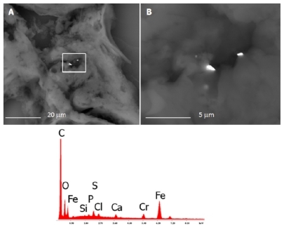
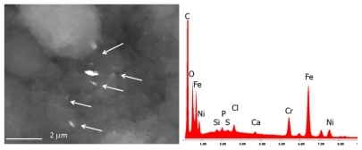
Figure 3 The bone marrow sample shows the presence of many nanoparticles (range 150 nm–5 μm) composed of iron, chromium, and nickel, namely debris of stainless steel.