Figures & data
Figure 1 Schematic overview of procedures used in this study.
Notes: (A, B) Isolation and expansion of MSCs from various tissues; (C) infection of MSCs with retrovirus carrying yCD∷UPRT suicide gene; (D) Selection of cell population of yCD∷UPRT gene-transduced cells; (E) labeling of yCD∷UPRT-MSCs with Venofer overnight; (F) preparation and harvesting conditional medium; (G) size characterization of yCD∷UPRT-MSCs/Venofer exosomes in comparison with Venofer; (H) treatment of tumor cells with CM from yCD∷UPRT-MSCs/Venofer cells and exposition to AMF; (I) determination of viability of tumor cells.
Abbreviations: 5-FU, 5-fluorouracil; AMF, alternating magnetic field; CM, conditioned medium; MSCs, mesenchymal stem cells; yCD∷UPRT, yeast cytosine deaminase∷uracil phosphoribosyl transferase suicide fusion gene.
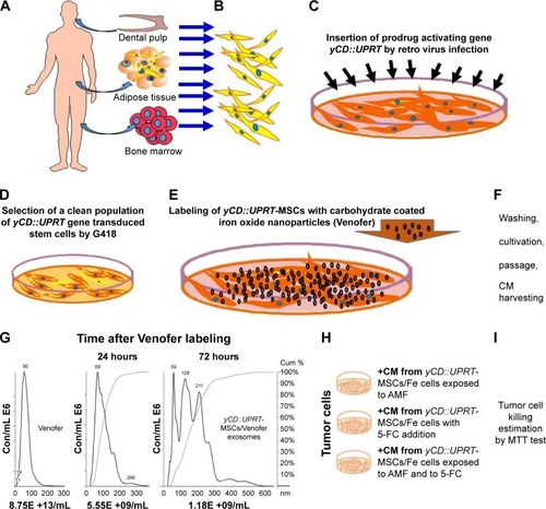
Figure 2 Growth of DP-MSCs/Fe and yCD∷UPRT-DP-MSCs/Fe cells in the absence and/or presence of 5-FC.
Notes: Kinetics of cell proliferation were measured in 96-well cell culture cluster (Corning) monitored by Incucyte system. Venofer was applied to the cell culture medium at concentrations of (A) 50 μg/mL, (B) 200 μg/mL, and (C) 400 μg/mL. (A–C) 5-FC (100 μg/mL) was added to each well with the CM 1 day after cell seeding. (D) Inhibition of proliferation of human prostate tumor cells following various doses of CM from yCD∷UPRT-AT-MSCs/Fe cells in the presence and/or absence of 5-FC. Concentration of 24 hour CM was quantified and expressed as μg of protein.
Abbreviations: 5-FC, 5-fluorocytosine; AT-MSCs, MSCs from adipose tissue; CM, conditioned medium; DP-MSCs, MSCs of the human dental pulp; MSCs, mesenchymal stem cells; yCD∷UPRT, yeast cytosine deaminase∷uracil phosphoribosyl transferase suicide fusion gene.
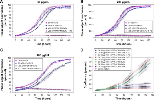
Figure 3 Characterization of nanoparticles released from DP-MSCs/Fe cells.
Notes: The media conditioned without PE for 24 hours by DP-MSCs/Fe cells labeled with various concentrations of Venofer were harvested. Media were centrifuged to remove cell debris and passed through a 0.2 μm syringe filter. The concentration and size distributions of nanoparticles in the CM of Venofer-labeled cells were measured with a NanoSight NS500 instrument. Prussian blue staining was used to detect iron in Venofer-labeled DP-MSCs.
Abbreviations: CM, conditioned medium; DP-MSCs, MSCs of the human dental pulp; PE, human platelet extract.
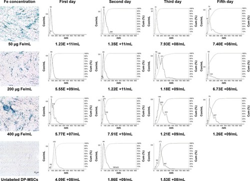
Figure 4 Kinetics of tumor cell death after AMF exposure following treatment with exosomes and secretome.
Notes: (A) Size-exclusion chromatography of CM harvested from yCD∷UPRT-DP-MSCs/Fe cells. The fractionation of 2 mL of CM on a Sephacryl 500 HR column was performed at 4°C. Each fraction was tested for stimulation of tumor cell proliferation in the absence of 5-FC and for proliferation inhibition activity in the presence of 5-FC using the Incucyte system and MTT assay. Iron content and the number of nanoparticles were quantified. (B) Kinetics of tumor cell death after AMF exposure. CM from BM-MSCs/Fe or UC-MSCs/Fe cells was added to PC3 tumor cells seeded in Petri dishes and exposed to an AMF for 20 minutes. Cell viability was estimated at 5 and 21 days of treatment. (C) Time course of internalization of magnetic nanoparticles by tumor cells. CM from yCD∷UPRT-DP-MSCs/Fe cells was added to PC3 tumor cells. At various time points (30, 60, 90, 120, and 150 minutes) after CM addition, cells were exposed to an AMF. Tumor cell viability was measured at 5 days using the MTT assay. *P<0.05, **P<0.01, ***P<0.001.
Abbreviations: 5-FC, 5-fluorocytosine; AMF, alternating magnetic field; BM-MSCs, bone marrow mesenchymal stem cells; CM, conditioned medium; DP-MSCs, MSCs of the human dental pulp; MSCs, mesenchymal stem cells; UC-MSCs, umbilical cord mesenchymal stem cells; yCD∷UPRT, yeast cytosine deaminase∷uracil phosphoribosyl transferase suicide fusion gene.
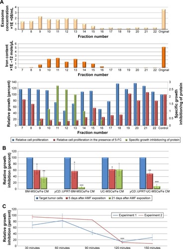
Figure 5 Kinetics of temperature increase for Venofer suspension in an AMF compared with control sample.
Note: Data obtained from three independent measurements are presented.
Abbreviation: AMF, alternating magnetic field.
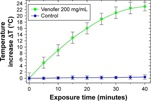
Figure 6 Representative examples of fiber-optic temperature profiles of exosomes incubated with different amounts of Venofer.
Abbreviations: CM, conditioned medium; MSCs, mesenchymal stem cells; yCD∷UPRT, yeast cytosine deaminase∷uracil phosphoribosyl transferase suicide fusion gene.
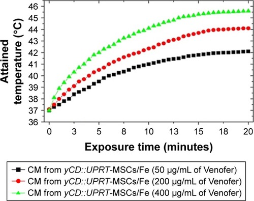
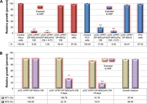
Figure 7 Effect of magnetic exosomes on tumor cells exposed to AMF.
Notes: (A) PC3 or HeLa cells were seeded into Petri dishes, and CM from yCD∷UPRT-DP-MSCs/Fe cells was added to the growth medium 1 day later with or without 5-FC. Cells were exposed to an AMF for 20 minutes. Five days after AMF treatment, cell viability was measured by the MTT assay. (B) Human primary glioblastoma (BT1) and recurrent astrocytoma (BT2) cells were seeded into Petri dishes, and CM from yCD∷UPRT-DP-MSCs/Fe cells was added to the growth medium 1 day later. Cells were exposed to an AMF for 20 minutes, and cell viability was measured 5 and 18 days using the MTT assay. **P<0.01, ***P<0.001.
Abbreviations: 5-FC, 5-fluorocytosine; AMF, alternating magnetic field; CM, conditioned medium; DP-MSCs, MSCs of the human dental pulp; MSCs, mesenchymal stem cells; yCD∷UPRT, yeast cytosine deaminase∷uracil phosphoribosyl transferase suicide fusion gene.