Figures & data
Figure 1 Synthesis and purification of RhoPro. (A) An NHS ester of Rho (1) reacts with N-terminal proline of Pro (2) to yield RhoPro (3). (B) High-performance liquid chromatography chromatograms show the separation of RhoPro from Pro and Rho. (C) Mass spectrometry showing four Pro components and four RhoPros generated by reaction of the N-terminal prolines as shown in A. (D) Absorption spectra of Pro, Rho, and purified RhoPro. Peaks 1–4 are Pro; peaks 5–8 are RhoPro.
Abbreviations: NHS, N-hydroxysuccinimide; Pro, protamine; Rho, rhodamine; RhoPro, rhodamine-protamine.
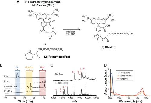
Figure 2 Uptake of RhoPro by HT-29 cells observed through FACS and the endosomal intracellular localization of RhoPro by fluorescence microscopy. (A) Dual-wavelength FACS scattergrams showing uptake of RhoPro and SYTOX Blue. (B) Time course of RhoPro uptake by single-channel FACS (0, 5, 15, 30, 60, and 120 min). Black line = 0 min, pink line = 5 min, lightest red line = 15 min, medium red line = 30 min, dark red line = 60 min, darkest red line = 120 min. (C) Median cell fluorescence from (B) plotted as a function of incubation time. Rho is rapidly internalized, while RhoPro is slowly internalized. (D) Intracellular localization of RhoPro on fluorescence microscopy. Rho fluorescence appears to be diffused throughout the cell. RhoPro colocalizes with LysoTracker Green (LTG), a marker of lysosomes. Magnification: 60× oil objective.
Abbreviations: DIC, differential interference contrast; FACS, fluorescence-activated cell sorting; fluores, fluorescence; Rho, rhodamine; RhoPro, rhodamine-protamine.
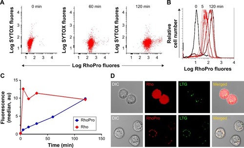
Figure 3 Phototoxicity of RhoPro-loaded HT-29 cells. (A) Dual-channel FACS scattergrams show RhoPro uptake (x-axis) and cell death (y-axis) with light and without light. Control groups indicate a role of irradiation only without any uptake. The blue circles indicate the positive fluorescence of the dead cell staining. (B) Data from the selected area of scattergrams are plotted as median fluorescence. The combination of light plus RhoPro uptake results in cell death. Microscopic imaging of light-induced cell death in (C) Rho- or (D) RhoPro-loaded cells. Cells were imaged by phase-contrast (DIC) microscopy with part of the field of view subjected to light. As shown in D, fluorescence of RhoPro decreased (bleached) while SYTOX Blue penetrated the illuminated cells. Magnification: 10× objective. Left side of the red dashed lines indicates regions with no light, while right side indicates regions with light.
Abbreviations: DIC, differential interference contrast; FACS, fluorescence-activated cell sorting; fluores, fluorescence; Pro, protamine; Rho, rhodamine; RhoPro, rhodamine-protamine.
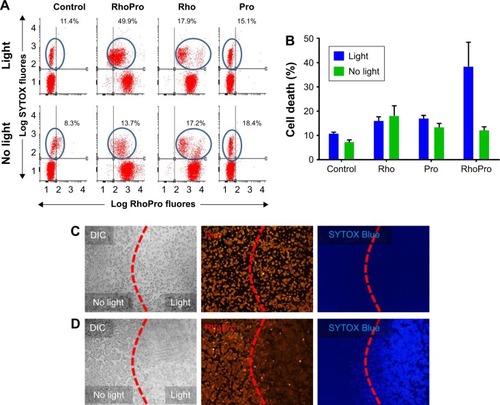
Figure 4 Kinetics of light-induced cell death in RhoPro-loaded cells. (A) With light, RhoPro fluorescence bleaches and induces RhoPro-loaded cells to take up SYTOX Blue. (B) Kinetics of light-induced RhoPro bleaching and SYTOX entry. Top two frames: DIC and Rho fluorescence shown as a function time of exposure to light. Bottom two frames: light induced SYTOX uptake. Magnification: 60× oil objective. (C) Time course for RhoPro and Rho bleaching and SYTOX uptake. RhoPro bleaches first, and cell death occurs as SYTOX entry commences. Additionally, the bleaching of rhodamine (Rho)-loaded cells and lack of SYTOX penetration are shown. (D) RNO bleaching assay showing generation of free oxygen radicals.
Abbreviations: DIC, differential interference contrast; fluores, fluorescence; Pro, protamine; Rho, rhodamine; RhoPro, rhodamine-protamine; RNO, N,N-dimethyl-4-nitrosoaniline.
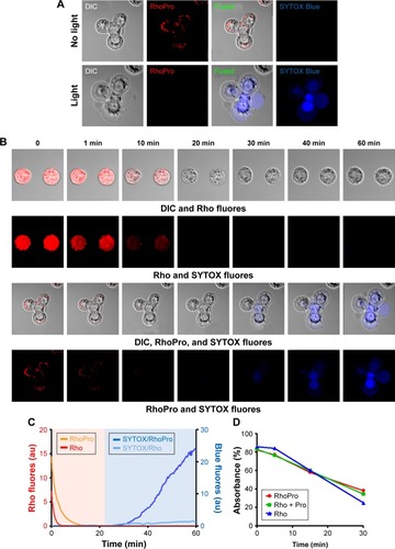
Figure 5 Lysosomal rupture induced by the photodynamic effect of RhoPro. (A) Internalized RhoPro was accumulated in lysosomes co-stained with the lysosome marker, LysoTracker Green. Yellow arrows indicate intact lysosomes. (B) After irradiation, lysosomal rupture occurred, and the fluorescence of LysoTracker Green and RhoPro decreased and appeared to diffuse in the cytoplasm. Magnification: 60× oil objective.
Abbreviations: DIC, differential interference contrast; RhoPro, rhodamine-protamine.
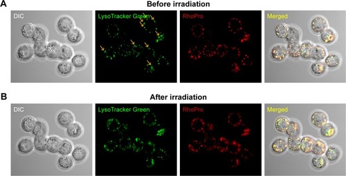
Figure 6 The in vivo photodynamic effect of RhoPro in tumor xenograft models. (A) Accumulation of RhoPro in tumor blood vessels confirmed with Alexa Fluor® 488 anti-mouse CD31 antibody. Magnification: 40× objective. (B) Biodistribution of RhoPro in HT-29 tumor-bearing mouse. The significant differences are shown as *P<0.05 and **P<0.01. (C) In vivo PDT of RhoPro compared with that of Rho. PDT by RhoPro suppressed the tumor growth rate.
Abbreviations: PDT, photodynamic therapy; Rho, rhodamine; RhoPro, rhodamine-protamine.
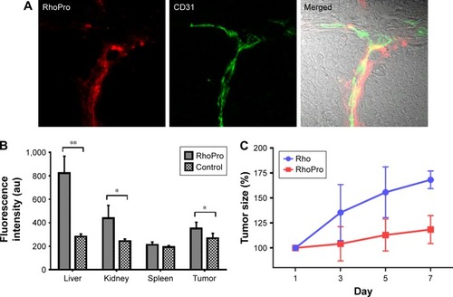
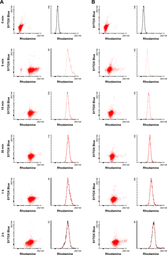
Figure S1 Cellular uptake and cytotoxicity of (A) RhoPro and (B) Rho in HT-29 cells determined through FACS analysis. Dual-wavelength FACS scattergrams showing uptake of the agents and cytotoxicity measured by the intensity of SYTOX Blue and time course of RhoPro uptake by single-channel FACS (0, 5, 15, 30, 60, and 120 min).
Abbreviations: FACS, fluorescence-activated cell sorting; Rho, rhodamine; RhoPro, rhodamine-protamine.