Figures & data
Figure 1 Types of synthesized nanocomposites.
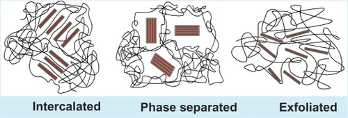
Figure 2 Different structures of silsesquioxanes: ladder structure (A), partial cage structure (B), and cage structure (C).

Figure 3 Biofunctionalization of the surface to enhance in situ endothelialization. Biofunctionalized surface of the heart valve leaflets made from POSS-PCU nanocomposite can target several biological processes to promote in-situ endothelialization, by promoting the mobilization of EPC from the bone marrow, encouraging cell-specific (circulating EC, EPC, and stem cells) homing to the vascular graft site, providing cell-specific adhesion motifs on the vascular grafts (of a predetermined spatial concentration), and directing the behavior of the cells post-adhesion to rapidly form a mature fully functioning endothelium with self-repair capability.
Copyright© 2008. American Chemical Society. Adapted with permission from De Mel A, Jell G, Stevens MM, Seifalian AM. Biofunctionalization of biomaterials for accelerated in situ endothelialization: a review. Biomacromolecules. 2008;9(11):2969–2979.Citation65
Abbreviations: EC, endothelial cell; EPC, endothelial progenitor cells; POSS-PCU, polyhedral oligomeric silsesquioxane-poly(carbonate-urea)urethane.
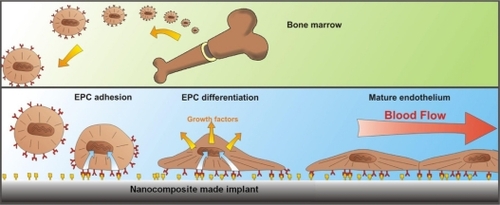
Figure 4 Morphological changes of isolated cells cultured on POSS-PCU nanocomposite polymer. Spindle-shaped morphology of early EPCs at day 7 (A) has been dominated by cobble stone-shaped features at day 21 (B) characteristics for the late EPCs or ECs; immunostaining of the cultured cells at day 14. Cells were stained for vWF (C) and VEGFR2 (D), showing positive expression of these cell surface markers on the cultured cells.
Abbreviations: EC, endothelial cell; EPC, endothelial progenitor cells; POSS-PCU, polyhedral oligomeric silsesquioxane-poly(carbonate-urea)urethane.
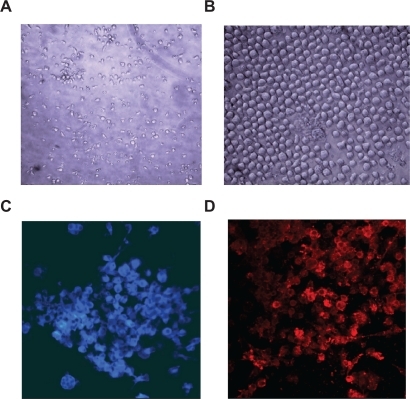
Figure 5 Chemical analysis of calcium deposition. Quantitative analysis of calcium deposition on the samples showed significantly reduced calcium deposition on POSS-PCU compared with BP (P < 0.001, n = 5) and in comparison with PU (P < 0.05, n = 5).
Abbreviations: BP, bovine pericardium; POSS-PCU, polyhedral oligomeric silsesquioxane-poly(carbonate-urea)urethane; PU, polyurethane.
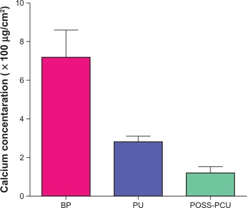
Figure 6 A) Trileaflet heart valve design (UCL design) with complex geometry and additional reflection on the leaflets to improve hemodynamic performance and durability. B) A valve prototype fabricated from POSS-PCU nanocomposite with a Dacron suture ring.
Abbreviations: POSS-PCU, polyhedral oligomeric silsesquioxane-poly(carbonate-urea)urethane; UCL, University College London.
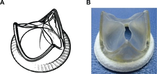
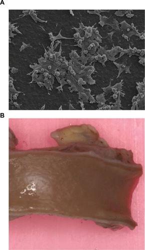
Figure 7 A) Scanning electron microscope pictures of endothelial cell adhesion morphology on POSS-PCU showing the presence of flat, spindle-shaped cells with numerous filopodia and the absence of cell retraction. This indicates the viability and proliferation of these cells on POSS-PCU at 48 hours (320× magnification). B) An explanted POSS-PCU bypass graft demonstrated to be endothelialized and patent after a 2 year implantation in a sheep model.
Abbreviation: POSS-PCU, polyhedral oligomeric silsesquioxane-poly(carbonate-urea)urethane.