Figures & data
Table 1 Grafting percentage of polymer on surface according to solvents used
Table 2 Grafting percentage of polymer on surface according to n-isopropylacrylamide concentration
Figure 1 A) Grafting percentage of n-isopropylacrylamide onto polystyrene with two different solvents and B) Effect of n-isopropylacrylamide concentration on grafting in 9:1 (v/v) water/methanol solvent.
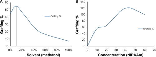
Figure 2 Attenuated total reflection Fourier transform infrared spectra of the ungrafted polystyrene (bottom) and spectra of the grafted polystyrene by ultraviolet radiation (top).
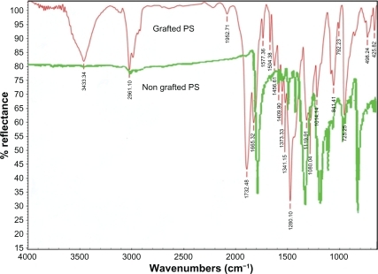
Figure 3 Atomic force microscopic image of grafted polystyrene under ultraviolet radiation (scale 1 × 1 μm). A) Preirradiated by gamma radiation (40 KGy), B) Grafted sample in water, C) Grafted sample in 9:1 (v/v) water/methanol solvent, D) Grafted sample in pure methanol solvent.
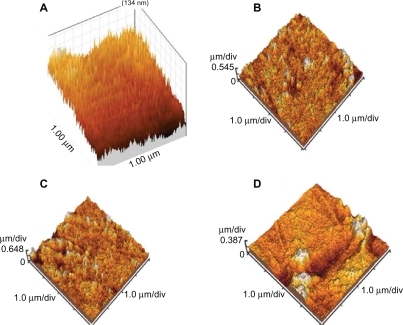
Figure 4 Atomic force microscopic image of grafted polystyrene with better grafting (120%) with 40% n-isopropylacrylamide concentration resolved in the solvent of 9:1 (v/v) water/methanol under ultraviolet radiation (scale 1 × 1 μm).
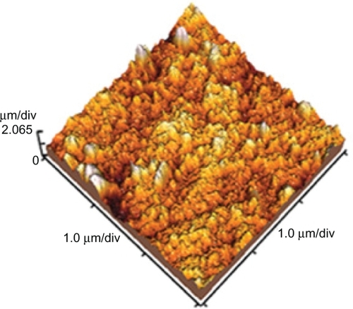
Figure 5 Scanning electron microscopy of grafted polystyrene under ultraviolet radiation. A) Grafted sample in water (cross section), B) Grafted sample in the solvent of 9:1 (v/v) water/methanol (cross section), C) Grafted sample in the solvent of 9:1 (v/v) water/methanol with 40% n-isopropylacrylamide (cross section), and D) Surface morphology of grafted sample.
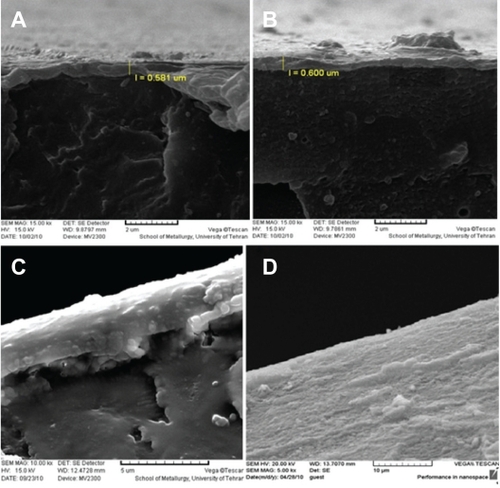
Table 3 Contact angles for normal and preirradiated and grafted samples
Figure 6 Differential scanning calorimetry spectra of the grafted polystyrene with 40% n-isopropylacrylamide concentration resolved in the solvent of 9:1 (v/v) water/methanol under ultraviolet radiation.
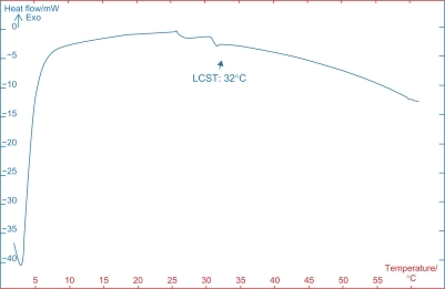
Figure 7 Cell viability in grafted sample with 40% n-isopropylacrylamide concentration resolved in a solvent of 9:1 (v/v) water/methanol under ultraviolet radiation at physiological temperature (37°C, ) and detached cells on the sample surface after 60 minutes of incubation at 4°C () with cell viability on control tissue culture polystyrene dishes.
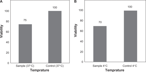
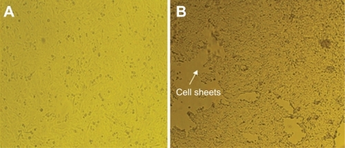
Figure 8 Epithelial cell growth on the grafted Petri dish at 37°C in , and in the cells detached spontaneously when temperature decreases below 4°C on the grafted sample is shown.